Figure 4. Protein expression of four selected genes in the cornea, limbus and conjunctiva by immunohistochemistry study. The left column
was cornea (A,D,G,F). The middle was limbus (B,E,H,K), and the right was conjunctiva (C,F,I,L). HOP (green, A-C) was localized to the nucleus (blue) of a subgroup of basal limbal epithelial cells (B, arrows) and stromal cells (arrow head). K13 (green, D-F) was expressed in the suprabasal limbal (E) and conjunctival (F) epithelium and absent in the cornea (C). PERP (green, G-I) was localized to all layers of the epithelium in all three tissues. TNC (green, J-L) was localized specifically at the basement membrane of the limbus and blood vessel walls (arrow).
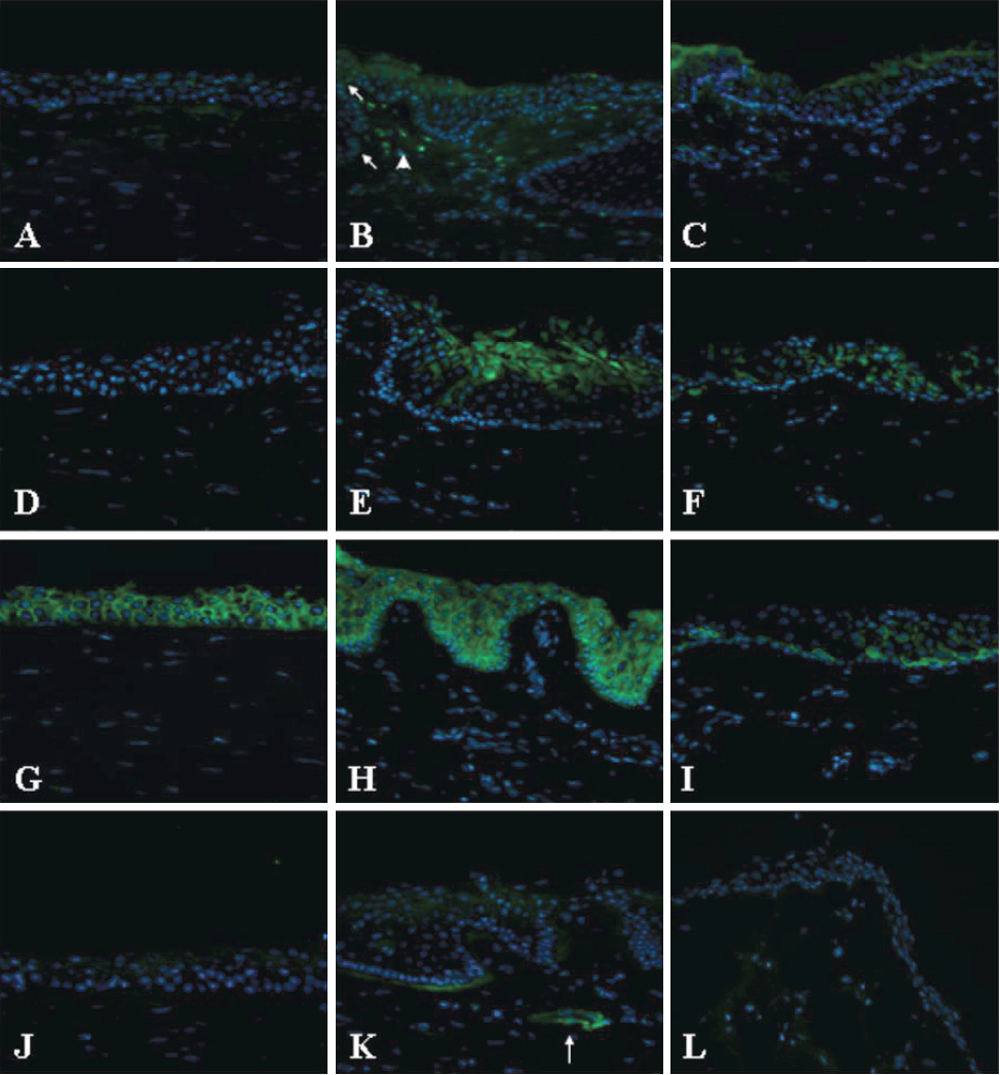
 Figure 4 of
Ding, Mol Vis 2008; 14:2031-2041.
Figure 4 of
Ding, Mol Vis 2008; 14:2031-2041.