Figure 2. Ultrastructural studies of
limbal, oral, and conjunctival cultures. A: The limbal
epithelial cell is shown with the desmosomes (D) and tight junction
(TJ) revealed in the inset. B: Oral epithelial cell is also
shown with the desmosomes (D) and gap junction (GJ) revealed in the
inset. C: Conjunctival epithelial cell is shown with just the
desmosomes (D) demonstrated in the inset.
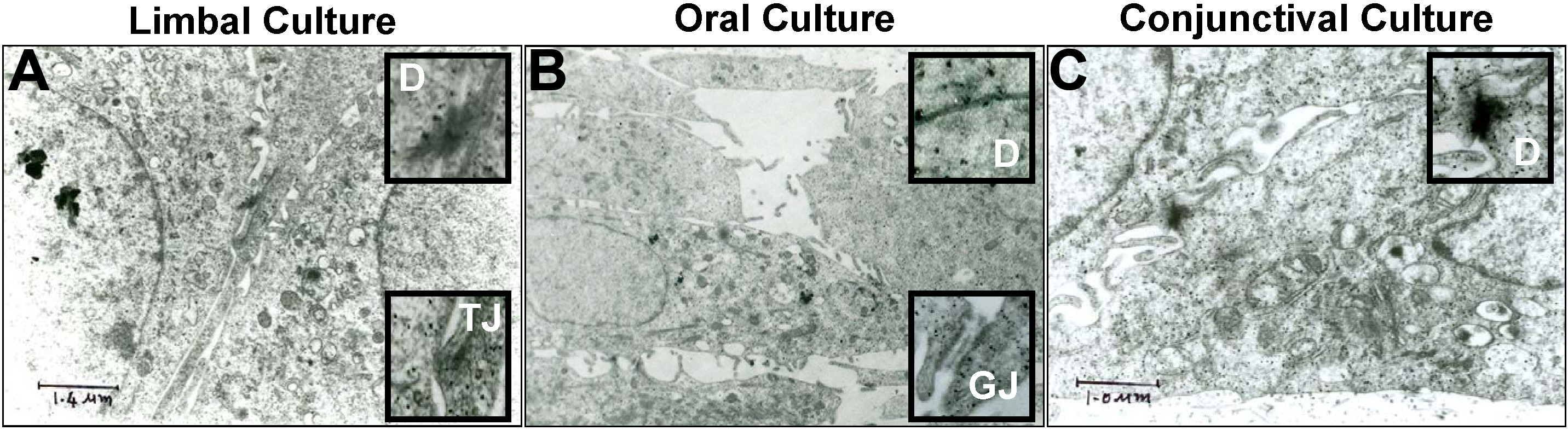
 Figure 2 of Madhira, Mol Vis 2008; 14:189-196.