Figure 1. Growth initiation and
morphological characteristics of limbal, oral, and conjunctival
cultures. A: Growth initiation from the three explant cultures
is shown. B: Confluent cultures of limbal, oral, and
conjunctival cells are shown. C: Hematoxylin and eosin staining
of sections of limbal, oral, and conjunctival cultures are illustrated.
D: PAS staining of the three explant cultures is also shown. The
inset in conjunctival culture shows goblet cells detected in native
conjunctival tissue.
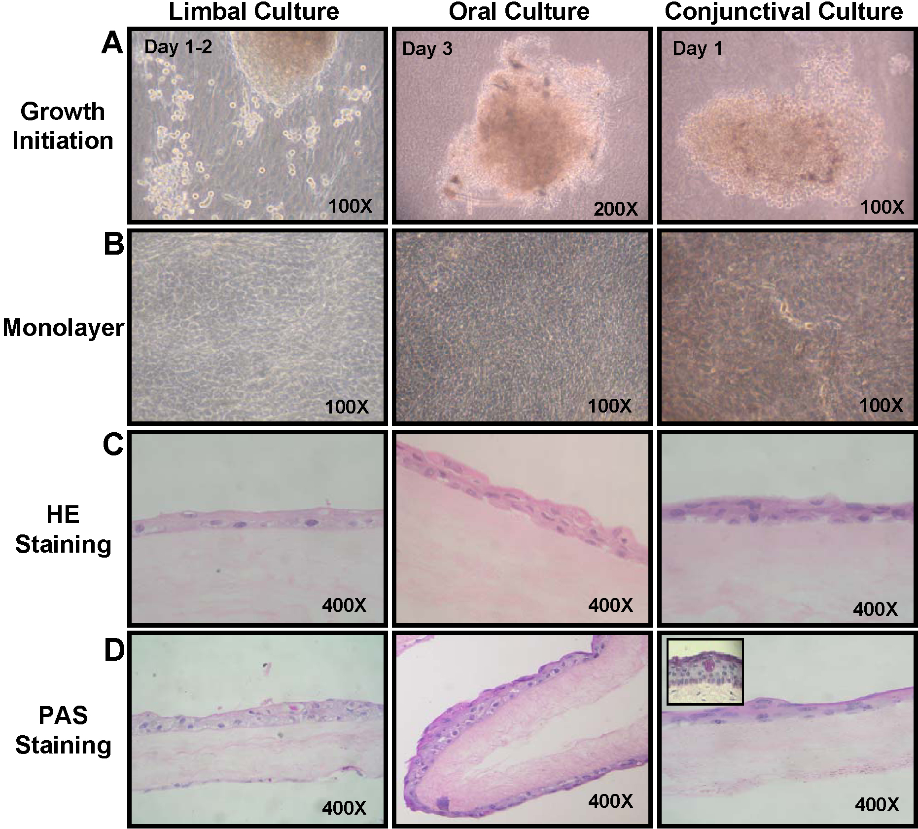
 Figure 1 of Madhira, Mol Vis 2008; 14:189-196.