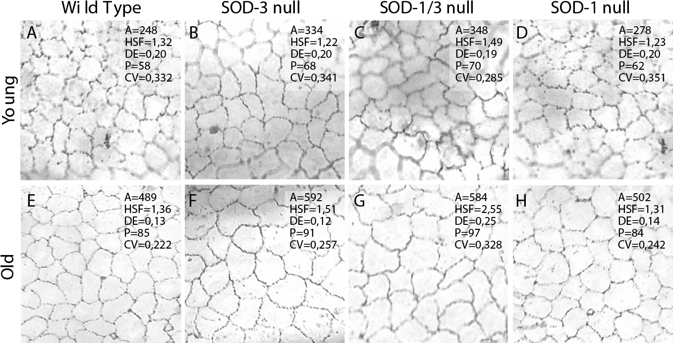
Figure 1. Corneal endothelial photographs.
A: Typical corneal endothelial photograph with alizarin red
staining from a C57BL/6 wild type mouse at a young
age. A=mean cell area, HSF=hexagon shape factor, quantifying
the deviation from the ideal hexagonal cell shape; DE=degree of cell
elongation; P=mean cell perimeter. B: SOD-3 null mouse,
young age. The cells are enlarged, compared to the wildtype control. C:
SOD-1/3 null mouse, young age. The cells are enlarged, compared to
the wildtype control. D: SOD-1 null mouse, young age. E:
Wild type mouse, old age. F: SOD-3 null mouse, old age. The
cells are enlarged, compared to the wildtype control. G:
SOD-1/3 null mouse, old age. Note that the cells are
enlarged and show increased pleomorphism and
polymegethism, compared to the wildtype control. H: SOD-1
null mouse, old age.