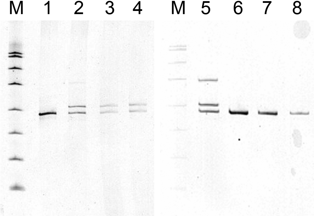
Figure 5. Prenatal diagnosis of the
inherited duplication. Gels of amplification products are shown. Lanes
1 and 6 are a negative control. Lanes 2 and 5 are the positive control,
III-5. Lanes 3 and 4 show two dilutions of the CVS material for IV-9.
Lanes 7 and 8 show two dilutions of the CVS material for IV-10. M is
the molecular weight marker, a HpaII digest of pUC19. IV-9 has the
insertion band and IV-10 has only the normal band.