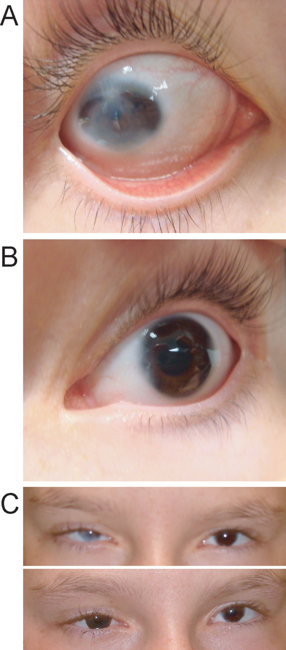
Figure 2. Proband, IV-2, at the age of 12
years and 6 months. A shows the severely affected right eye
with peripheral sclerocornea. B shows the mildly affected left
eye, with peripheral corneal stromal opacity and displaced
Schwalbe's line. C shows the proband's eyes without (upper) and
with (lower) a colored contact lens in the right eye to mask the
corneal opacity.