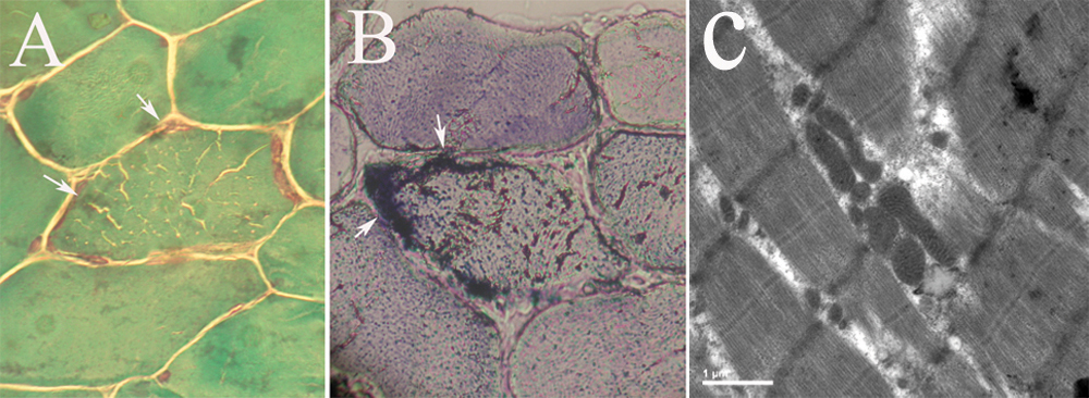
Figure 3. Light microscopic examination
and electron microscopic examination of a 10 μm skeletal muscle
transverse section. Gomori’s trichrome stain showed possible red ragged
fiber (RRF; A). Succinate dehydrogenase (SDH) staining showed
enhanced staining of the fiber edges (see arrows in B).
Electron microscopic examination showed mitochondrial proliferation and
enlarged mitochondria in a few muscle fibers, there were no crystalloid
inclusions (C).