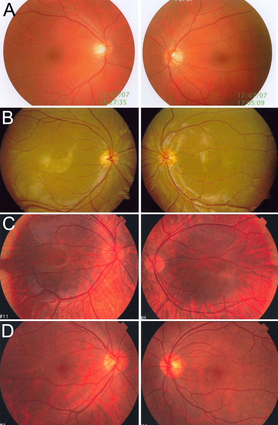
Figure 3. Photographs of fundi from normal
control, patients, and carriers. Both of eyes′ fundi are presented. A
is the fundi from a normal control. B shows the fundi of
proband (IV:16) in family 1. There is normal pigmentation and severe
foveal hypoplasia in both of eyes in picture B. C
presents the fundi of proband IV:1 from family 4. There was
hypopigmentation in the posterior of the fundus and severe foveal
hypoplasia in both of eyes in picture C. D shows the
fundi from carrier III:3 in family 4. There was pigmentary mosaicism in
the retinal pigment epithelium, and the fovea was normal in both of
eyes.