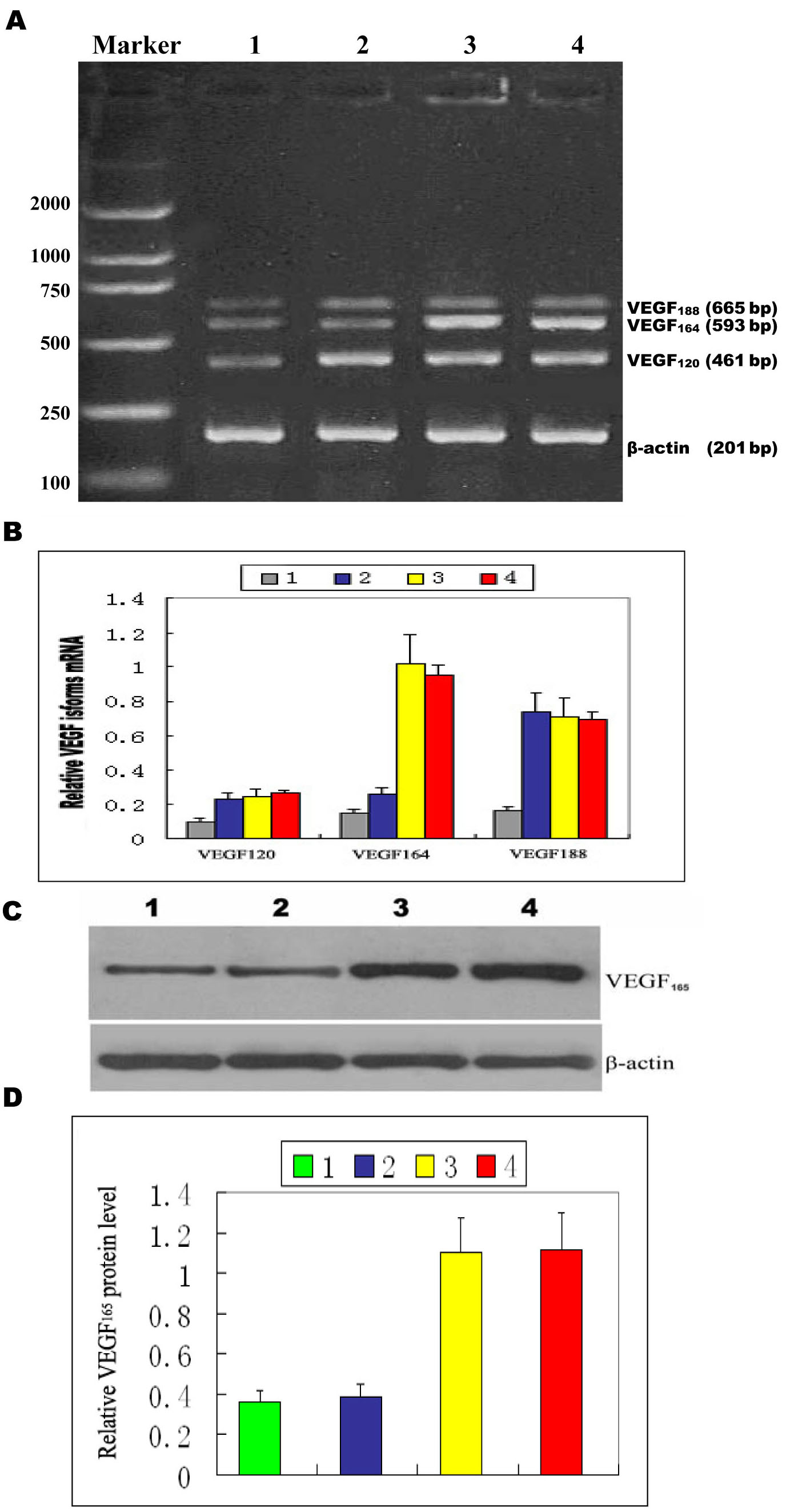
Figure 5. Suppression of vascular
endothelial growth factor expression in murine retinas by pSilencersiVEGF.
Murine retinas were extracted and examined for VEGF level. One-way
ANOVA followed by the LSD-t-test were used to evaluate
significant differences. A p value <0.05 was considered
statistically significant. Error bars are standard error of mean (SEM).
A: VEGF120, VEGF164, and VEGF188
mRNA levels in murine retinas were examined by RT–PCR. VEGF164
mRNA in murine retinas was downregulated after intravitreal
administration of pSilencersiVEGF, whereas VEGF120
and VEGF188 levels in murine retinas remained unchanged.
Lanes are marked as follows: 1) room air-raised mice; 2) murine model
with pSilencersiVEGF injection; 3) murine model with
pSilencer null vector injection; and 4) murine model of oxygen-induced
retinopathy. B: Relative VEGF isoforms mRNA quantification was
related to β-actin mRNA. C: Shown is an immunoblot assay for
VEGF165 protein in the murine retinas. The total amount of
VEGF164 immunosignal in murine retinas was reduced by
pSilencersiVEGF. The following abbreviations are used in
panel C: ganglion cell layer (GCL), inner nuclear layer (INL),
and outer nuclear layer (ONL). D: The levels of VEGF165
were quantitated by densitometry and normalized to β-actin protein
levels. Data are expressed as mean±SEM and were analyzed by one-way
ANOVA followed by LSD-t-test. The means of groups 3 and 4 are
statistically greater than the means of groups 1 and 2 (p<0.05).