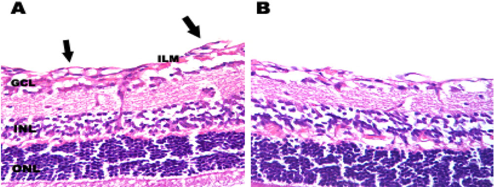
Figure 3. Histological analysis of the
effects of treatment with pSilencersiVEGF on
ischemia-induced retinal neovascularization. A: Postnatal day
17 (P17) retina from the eye of a hypoxic mouse injected with 1000 ng
pSilencer null vector. Extensive preretinal neovascular loops are
apparent (black arrows). B: P17 retina from the eye of hypoxic
mouse injected with 1000 ng pSilencersiVEGF. Preretinal
neovascular loops were not as apparent, suggesting that treatment
reduced preretinal neovascularization compared with the pSilencer null
vector-injected eye. The following abbreviations were used in the
figure: interlimiting membrane (ILM), ganglion cell layer (GCL), inner
nuclear layer (INL), and outer nuclear layer (ONL).