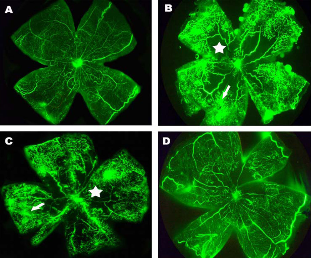
Figure 2. Angiographic analysis of the
effect of pSilencersiVEGF on the murine model of retinal
neovascularization. Retinal flatmounts were examined by fluorescein
dextran angiography. A: Shown is a retinal flatmount of mice
raised in room air. B: In this retinal flatmount of mice
exposed to hyperoxia, neovascular tufts appear as hyperfluorescence at
the junction between the perfused and the nonperfused area. Arrow
points to retinal neovascularization, and star marks an area of
nonperfusion. C: In this retinal flatmount of hyperoxia exposed
mice injected with pSilencer null vector, retinal neovascularization
and nonperfusion area were not attenuated. D: Shown is a
retinal flatmount of hyperoxia-exposed mice injected with pSilencersiVEGF.
The amount of neovascular tufts was markedly reduced and the area of
nonperfusion was diminished.