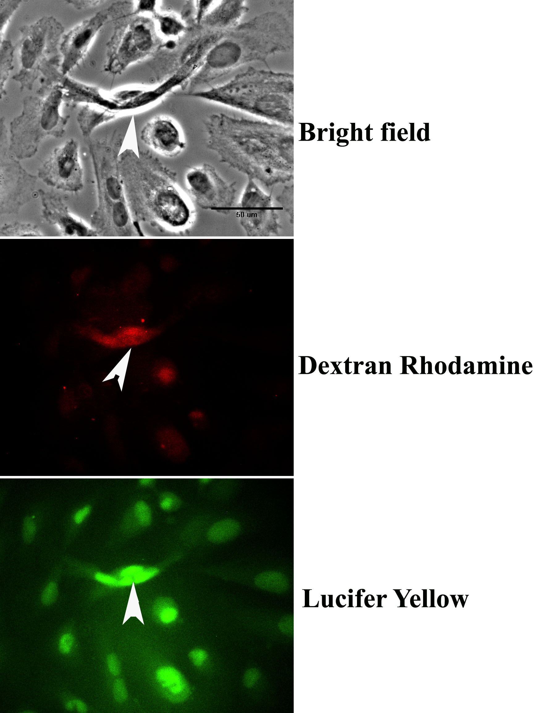
Figure 2. Dye spread in electrically
coupled cells. Gap junctions mediate the spread of Lucifer yellow (LY),
indicating that junctional permeability and the system of intercellular
communication are intact. Arrowheads in all panels show the location of
the small injury (scrape) produced by the tip of the scalpel blade.
Fluorescent images of cells incorporating both dextran rhodamine (DR)
and LY show that whereas the rhodamine complex did not spread from the
site of the scrape (middle panel), LY spread through the gap junctions
that coupled the cells (lower panel). Scale bar (upper panel) equals 50
μm.