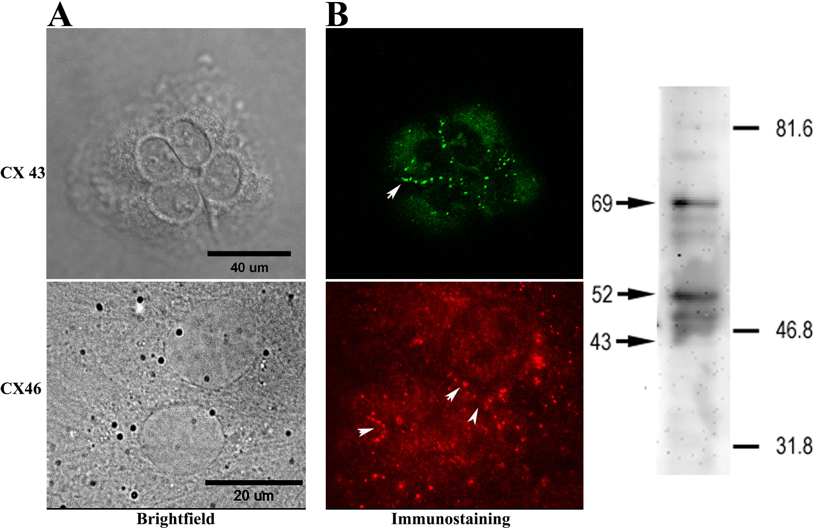
Figure 1. Connexin expression in ARPE-19
cells. A: Immunocytochemistry: an antibody to connexin 43 was
revealed with Alexa 488-tagged secondary antibody, and the antibody to
connexin 46 was visualized by fluorescent microscopy with a cy3-tagged
secondary antibody. Punctate labeling characteristic of connexin
plaques was seen both in the region of cell apposition (arrows) and at
the cell surface. In the left panel, scale bar in the Differential
interference contrast (DIC; upper) image represents 40 μm, while scale
bar in the phase (lower) image represents 20 μm. B: Western
blotting: Aliquot of whole cell ARPE-19 lysate (20 µg total protein)
were resolved by SDS–PAGE on a 6% Laemmli gel and transferred to PVDF
membrane. Anti-Cx43 antibody labeled several bands corresponding to
phosphorylated species of Cx43 (major bands identified by arrows at 69
and 52) and a minor band corresponding to nonphosphorylated Cx43 (arrow
at 43). Numbers and lines to the right indicate the positions of
molecular weight markers.