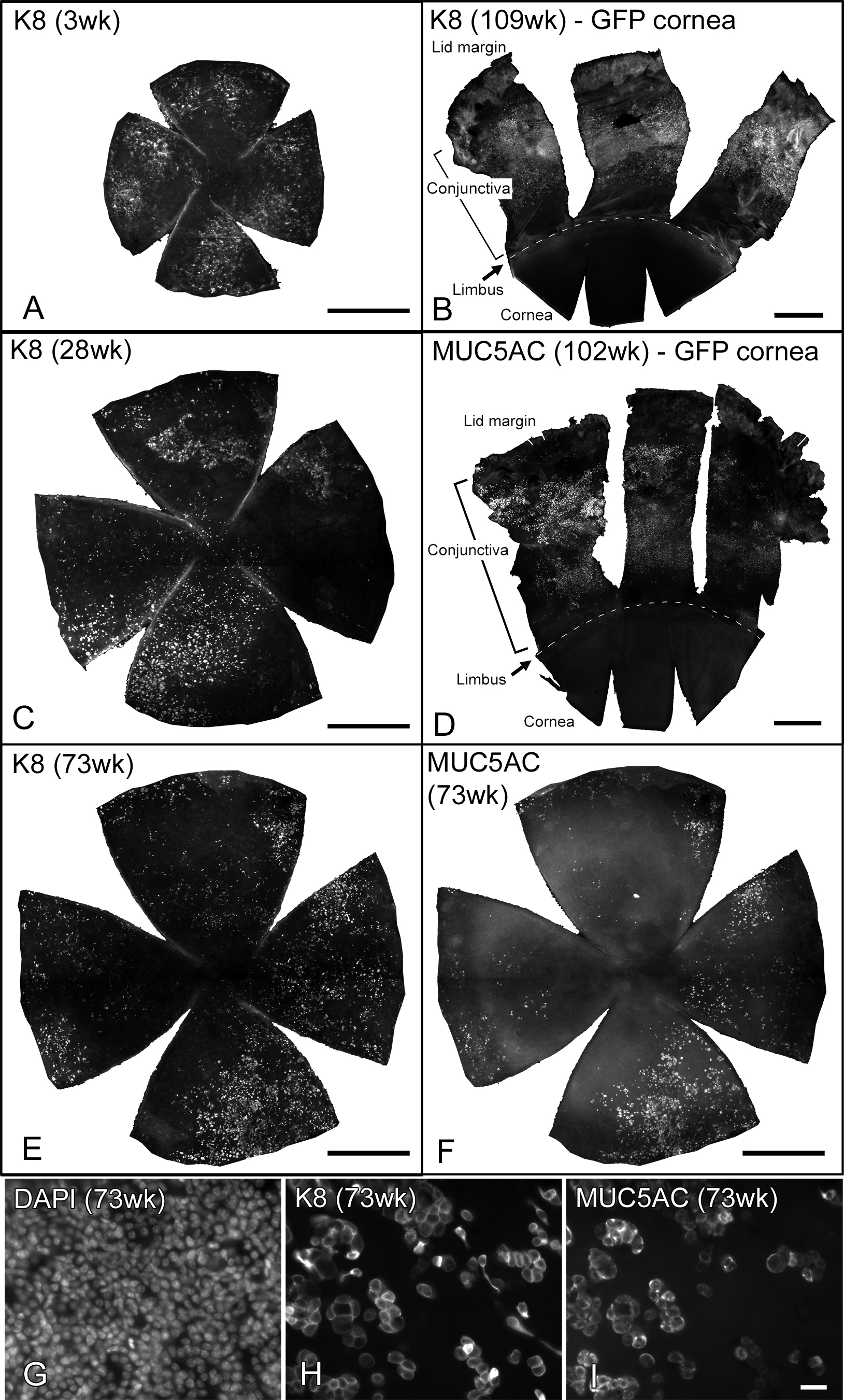
Figure 7. Expression of K8 and MUC5AC.
Immunofluorescence staining with K8 (A-C,E,H)
and MUC5AC (D,F,I) was performed with ocular
surface whole mounts of GFP-Dstncorn1 mice (A,C,E-I)
and CAG-EGFP mice (B,D) at the indicated ages. For the
GFP-Dstncorn1 specimens, only the cornea is shown,
and for the CAG-EGFP specimens, a superior half of the ocular surface
containing cornea and conjunctiva up to the lid margin is shown
together with an approximate position of the cornea-limbus boundary
line shown as dashed lines. B and D show that K8 and
MUC5AC are completely absent in normal CAG-EGFP corneas even at an
advanced age while they are present in wide areas of the conjunctiva. A,
C, and E show that K8 is widely distributed in GFP-Dstncorn1
corneas at all ages. E and F show the same GFP-Dstncorn1
cornea (73 weeks old) with double staining with K8 and MUC5AC,
respectively. G, H, and I show high power
images of the same 73-week-old cornea with triple staining with DAPI,
K8, and MUC5AC. Most of the MUC5AC positive cells are also positive
with K8. Bars: 500 µm (A-F), 20 µm (G-I).