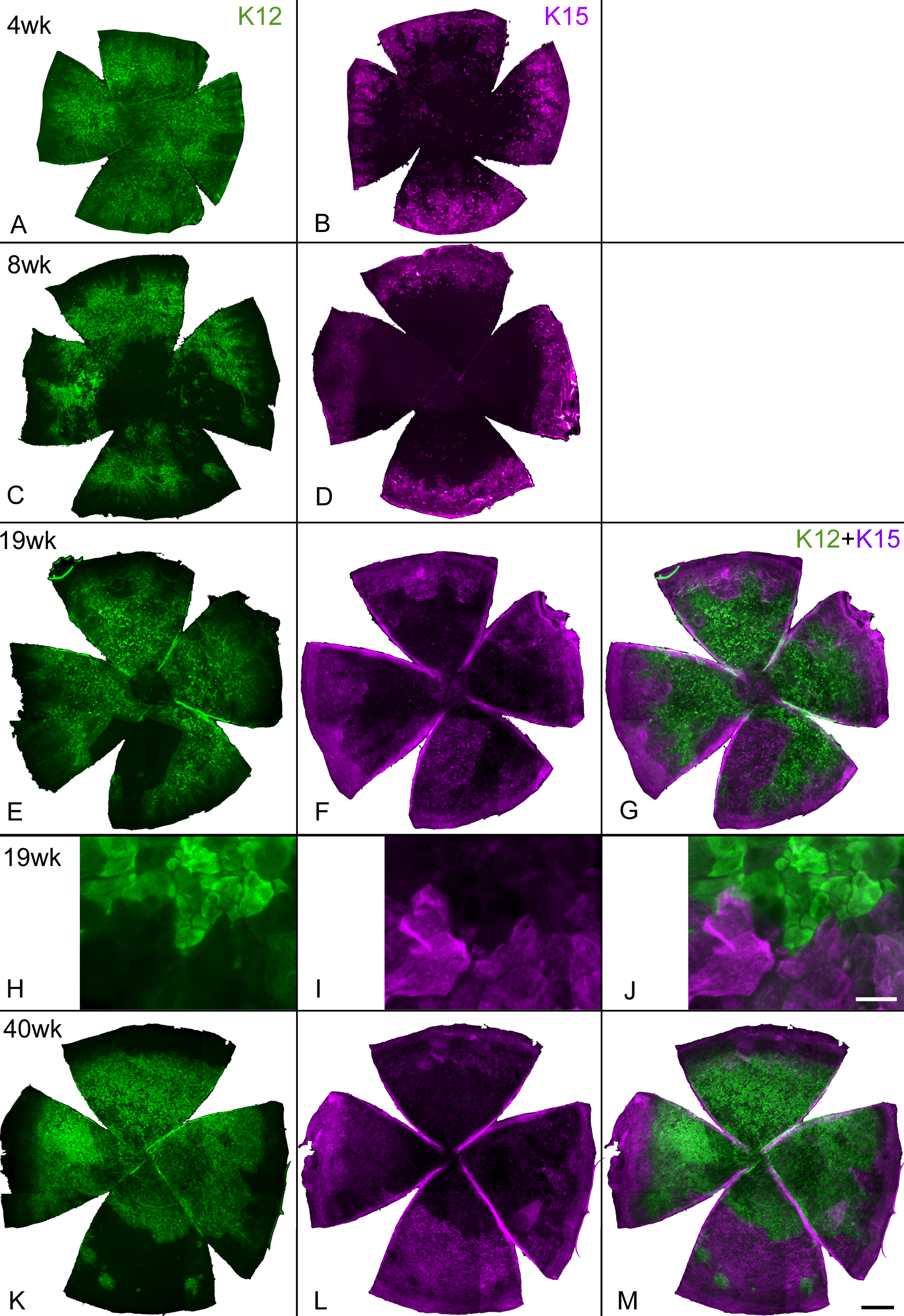
Figure 6. Expression of K12 and K15 in GFP-Dstncorn1
corneas at different ages. Whole mounts of GFP-Dstncorn1
cornea were used for immunofluorescence staining with K12 (A,C,E,H,K
in green) and K15 (B,D,F,I,L in
purple) at indicated ages. Only the cornea is shown. K12 patterns
varied in different corneas, but they were present in wide areas of the
GFP-Dstncorn1 cornea at all ages examined. K15
staining was present in a wide peripheral zone of the cornea at four
weeks (B), and the positive areas appeared to increase in older
corneas (D,F,L) while it was only present at a
very narrow peripheral zone of CAG-EGFP corneas adjacent to the limbus
at all ages (not shown). E and F, H and I,
and K and L are double staining of the same corneas,
and corresponding composite images of K12 and K15 are also shown (G,J,M).
A-D are all different corneas. In both a 19-week-old
cornea (E-G for low power, H-J for high
power) and a 40-week-old cornea (K-M), cellular patterns
of K12 and K15 are mostly complementary and exclusive, although the
match is not always perfect. Bars: 500 µm (A-G,K-M);
20 µm (H-J).