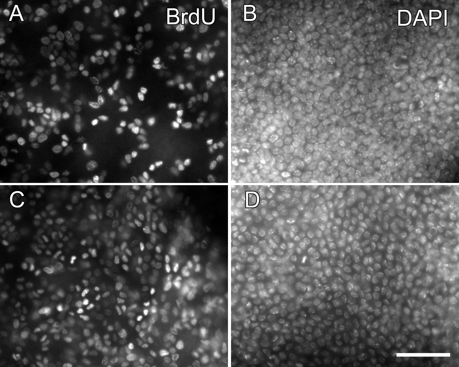
Figure 4. Distribution of mitotically
active cells. DNA synthesis was
determined in a normal 18-week-old
CAG-EGFP cornea (A,B) and a 20-week-old
GFP-Dstncorn1 cornea (C,D). Animals were labeled
with systemic BrdU for 24 h continuously with an osmotic pump. BrdU
positive cells were determined by immunohistochemistry with whole mount
corneas and basal epithelial cells imaged (A,C). DAPI
stain shows the entire population of basal epithelial cells of the same
area (B,D). Bar: 50 µm.