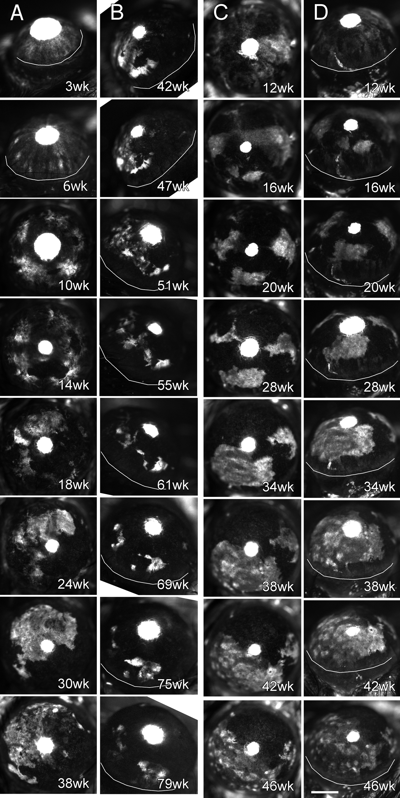
Figure 3. Two representative time lapse
sequences of corneal GFP in GFP-Dstncorn1 mice. One
representative time lapse is shown A and B and the
other in C and D. Corneal GFP patterns were imaged with
in vivo fluorescence microscopy at an interval of one to four weeks,
and selected images are shown together with the age of the mouse. White
areas represent GFP positive epithelial cells. Small central white
circles are due to GFP in the lens that was visible through a pupil and
masked corneal GFP. A: A time lapse sequence of central corneal
images of a GFP-Dstncorn1 mouse from six weeks to 42
weeks of age is shown. B: The same eye is shown as in the
previous panel (A), but a temporal side view from 42 weeks to 79
weeks is shown. C: A time lapse sequence of central corneal
images of another GFP-Dstncorn1 mouse is shown. D:
The same eye is shown at the same time points as in the latter panel (C)
except that images were from a temporal side view. White curved lines
are drawn to indicate a boundary between the cornea and the limbus in
panels with an angled view. Bar: 1 mm.