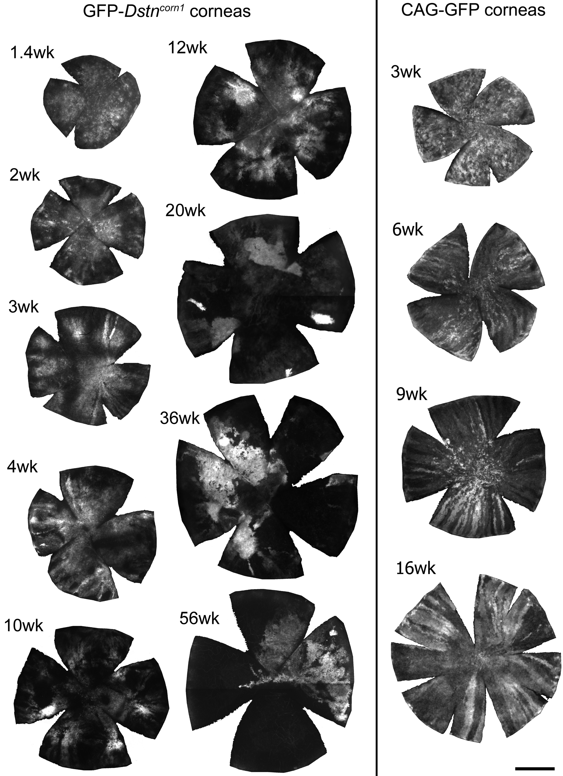
Figure 2. Developmental changes of GFP
patterns in GFP-
Dstncorn1 corneas and normal CAG-EGFP
corneas. Flat whole mount corneas were prepared from respective mice of
indicated ages. Cuts were made to mount the cornea flat. GFP patterns
were imaged and digitally recorded. Only the cornea area is shown.
Radial GFP stripes appeared in normal CAG-EGFP corneas around four
weeks and remained in all corneas of all ages we observed (see also [
10]). GFP stripes were
detected in young GFP-
Dstncorn1 mice at three to six
weeks, but there were none in adult corneas, which instead showed
globular and diffuse GFP patterns. Bar: 1 mm.