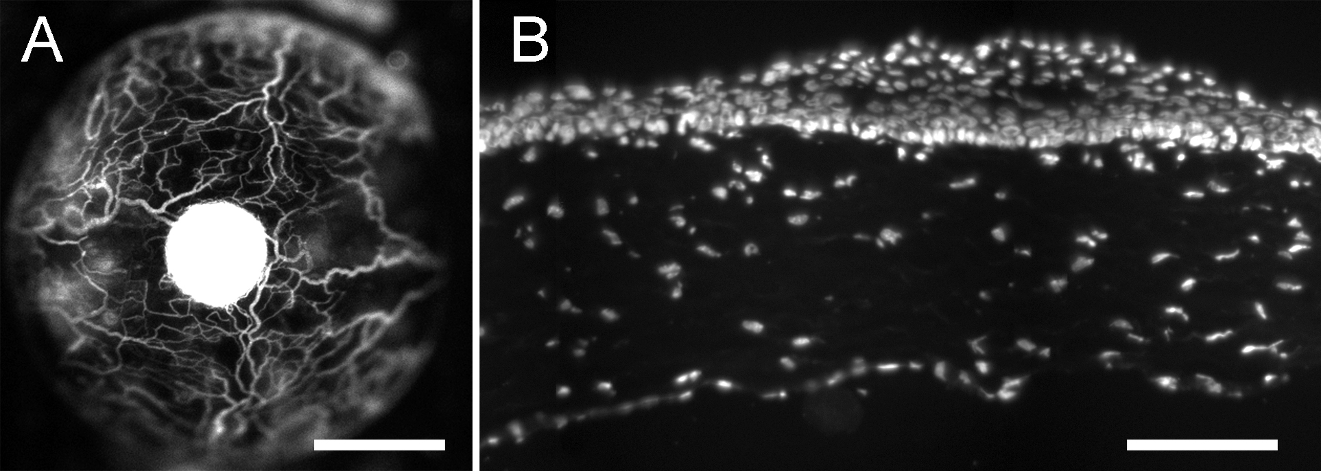
Figure 1. Expression of Dstncorn1
phenotype by GFP-Dstncorn1 corneas. A: The
corneal vasculature was visualized by sulforhodamine-101 angiography
with a 15-week-old GFP-Dstncorn1 mouse. B: A
cryosection of a cornea from a 12-week-old GFP-Dstncorn1
mouse was stained with DAPI, showing epithelial hyperplasia. Bar: 1 mm (A),
100 µm (B).