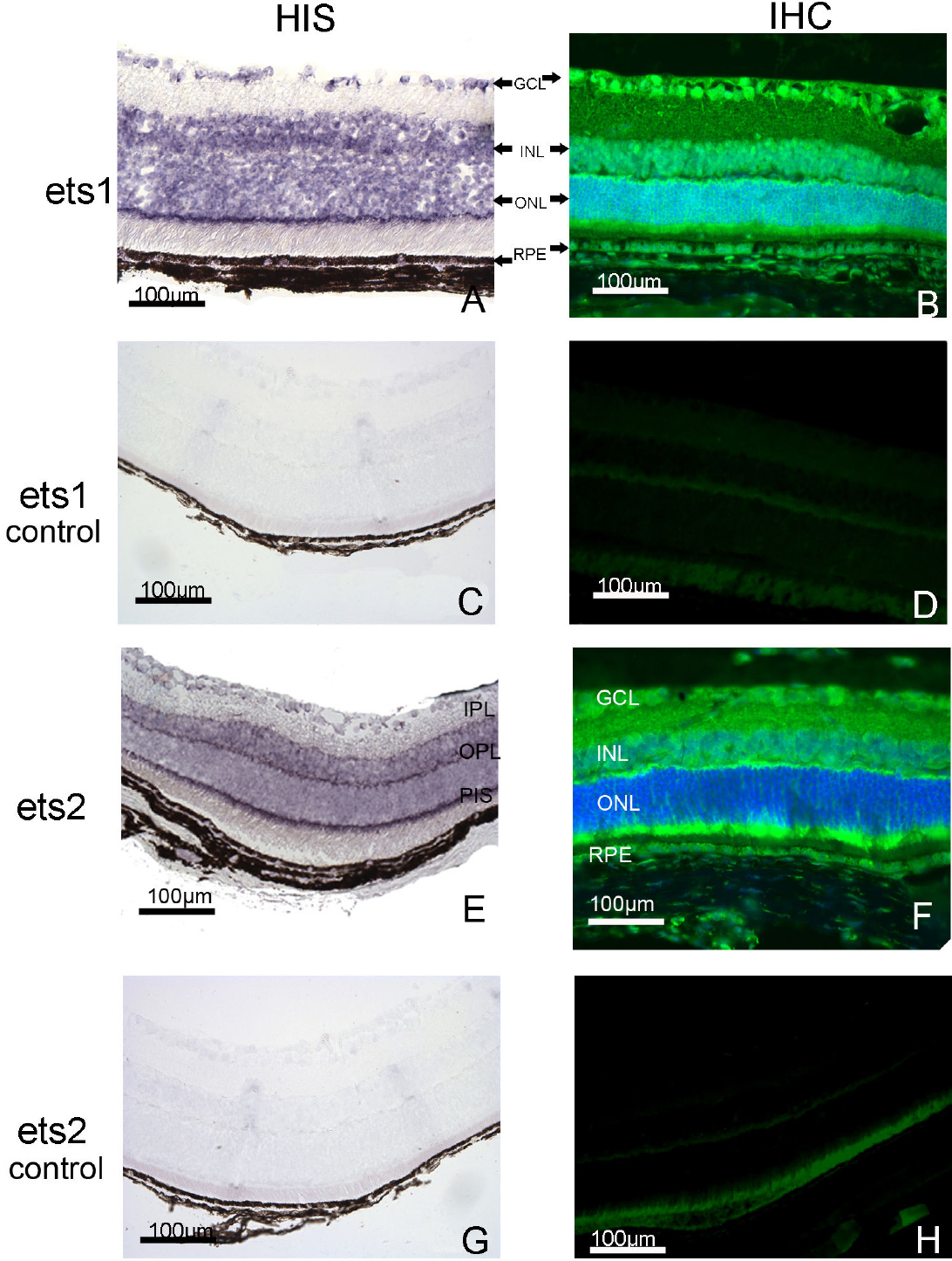
Figure 1. Distribution of ETS-1
and ETS-2 mRNAs and proteins in the adult mouse eye. The ETS-1
and ETS-2 mRNAs were detected in adult mouse tissue sections by
in situ hybridization (HIS), and their distributions are shown (A
and E, respectively). The ETS-1 and ETS-2 proteins were
detected by immunohistofluorescence (IHC) in adult mouse tissue
sections. The specific immunostaining patterns obtained are also shown (B
and F, respectively). The retinal tissue distributions of ETS-1
(B) and ETS-2 (F) proteins appeared to be somewhat
different whereas the distribution of the transcripts corresponding to
these proteins (A and E) seemed to be similar in adult
mouse retina. The ETS-1 (C)and ETS-2 (G)
mRNAs show the negative control in adult mouse tissue sections by in
situ hybridization obtained with sense riboprobes. The ETS-1 (D)
and ETS-2 (H) immunohistofluorescence shows the negative control
in adult mouse tissue section. The results were obtained with the
second antibody alone. The nucleus was counterstained with DAPI. The
scale bar represents 100 µm. GCL: ganglion cell layer; IPL: inner
plexiform layer; INL: inner nuclear layer; OPL: outer plexiform layer;
ONL: outer nuclear layer, RPE: retinal pigment epithelium; PIS:
photoreceptor inner segment.