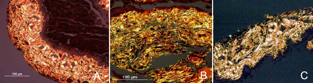
Figure 2. Analysis of iris sections by
Sirius red staining. Iris sections from PACG (A), POAG (B)
as well as normal (C) patients were stained with Picrosirius
red. When viewed with a polarized light, mature type I collagen fibers
appear bright yellow or orange. Magnification x20.