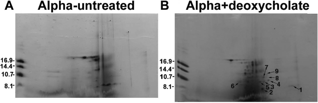
Figure 2. Proteolysis of αA- and
αB-crystallins following incubation of the α-crystallin fraction with
sodium deoxycholate at 37 °C for 10 h. Deoxycholate untreated
α-crystallin fraction (A) and deoxycholate (2% w/v)-treated
α-crystallin fraction (B; each containing 190 μg protein at
37 °C for 10 h) were analyzed by two-dimensional gel
electrophoresis, and those spots present in only the
deoxycholate-treated preparations were identified and numbered as 1-9
as shown in (B). Each spot was analyzed by the MALDI-TOF mass
spectrometric method to determine their identity.