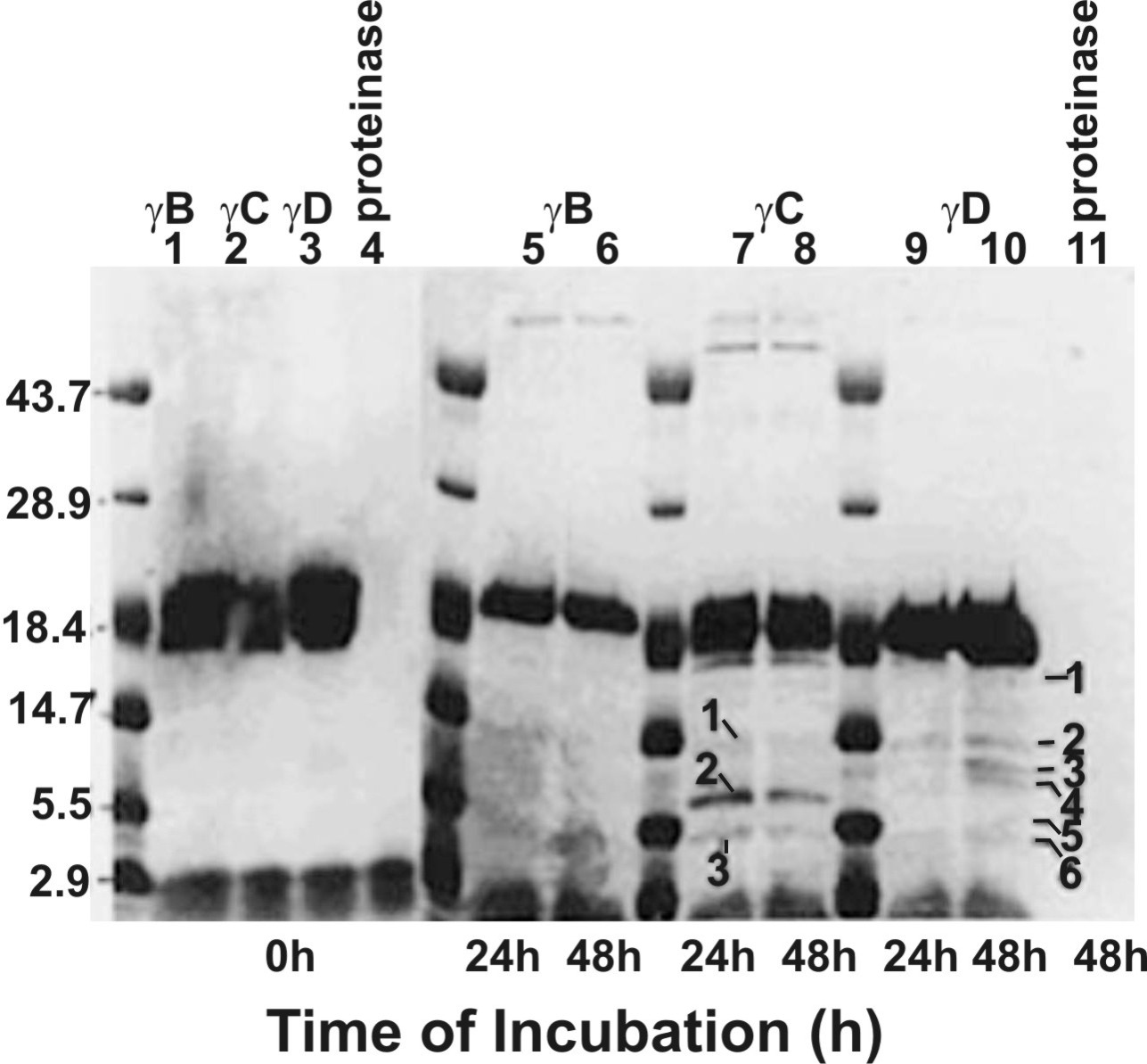
Figure 10. Time-dependent proteolysis of
bovine recombinant γB-, γC-, and γD-crystallins by the human lens
membrane proteinase. The incubation periods are shown at the bottom of
the gel, and the reaction constituents are shown at the top of the gel.
The proteolyzed fragments are identified with numbers, and only the
fragments from γD-crystallin were used for partial NH2-terminal
sequence analysis.