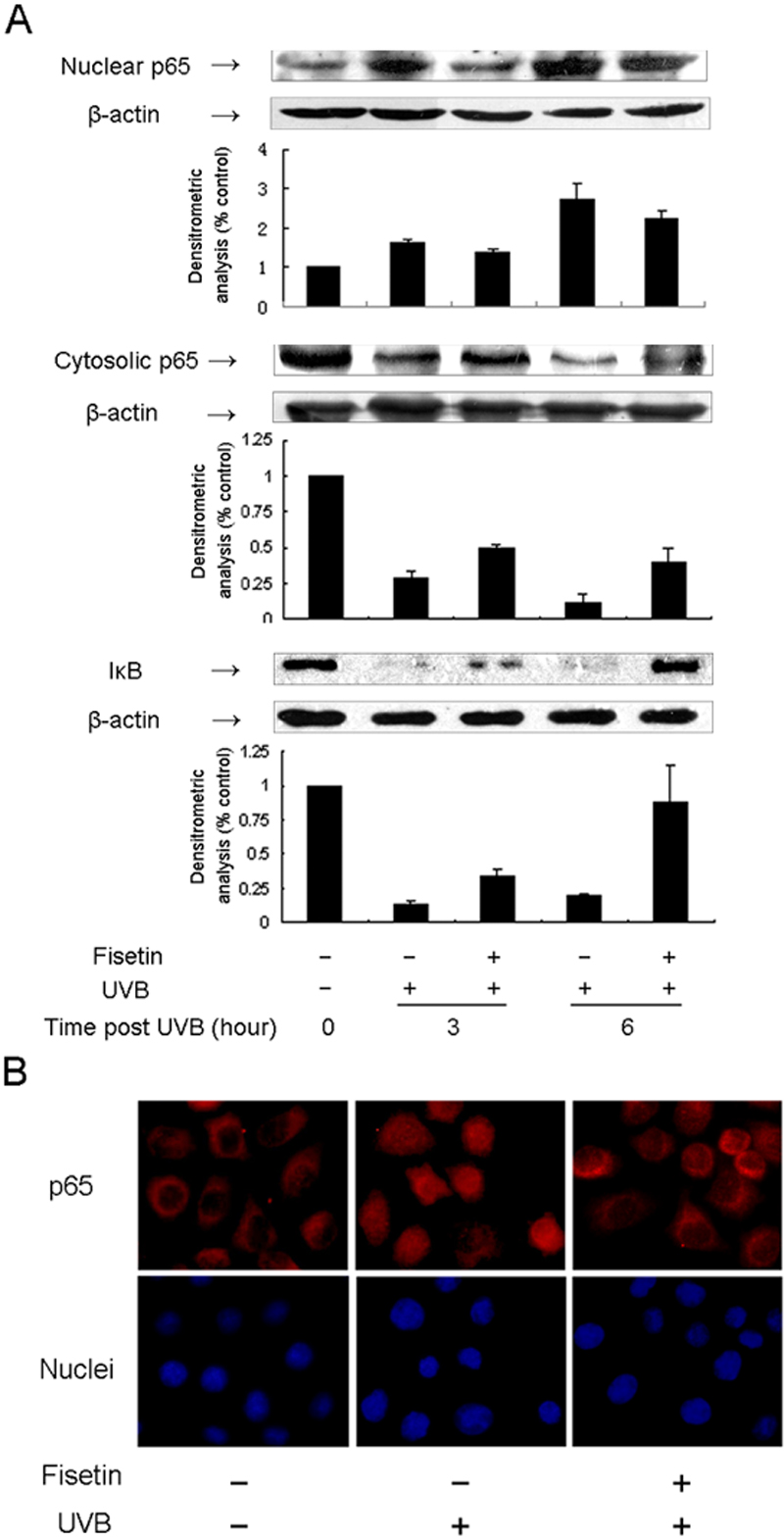
Figure 3. Effect of fisetin on UVB-induced
activation of NF-кB and degradation of IкB in HLE cells. A:
SRA01/04 cells were exposed to UVB (30 mJ/cm2) with or
without pretreatment with fisetin (25 μg/ml) for 1 h. Cells were
harvested at 3 h and 6 h time points after UVB exposure, and cell
lysates were prepared to determine the activation of NF-кB or
degradation of IкB using western blot analysis. The graph represents
the quantification results normalized to β-actin levels. Data represent
the mean±SD of three individual experiments. The asterisk indicates
p<0.05. B: Immunocytochemical analysis of NF-кB p65
localization is visualized. Cultured cells were incubated with anti-p65
antibody overnight at 4 °C as described in Methods. p65 is stained
red, and the nuclei are stained blue. Representative fluorescent images
were taken under fluorescence microscopy. Magnification, 100X.