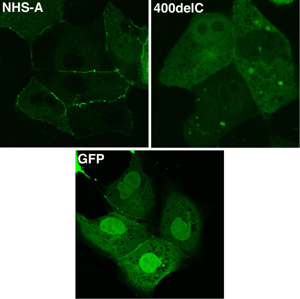
Figure 3. Localization of GFP-NHS-A400delC
mutant in MDCK cells. Cells were transfected with GFP-NHS-A and
GFP-NHS-A400delC fusion constructs and pEGFP-C1 control. Transiently
expressed fusion proteins were visualized by confocal microscopy.
GFP-NHS-A wild type protein primarily localized to the cellular
periphery whereas GFP-NHS-A400delC mutant protein localized in the
cytoplasm and nucleus. Apparent peripheral distribution of GFP is an
experimental artifact seen only between some adjoining transfected
cells. Images were taken with a 60X objective.