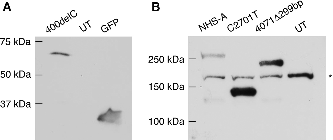
Figure 2. Expression of mutant NHS-A
proteins in mammalian cells. A: Lysates of HEK 293A cells
transiently transfected with GFP-NHS-A400delC and pEGFP-C1 constructs
and untransfected cells were analyzed by western blotting with anti-GFP
antibody. B: Lysates of HEK 293A cells transiently transfected
with FLAG-NHS-A, FLAG-NHS-AC2701T and FLAG-NHS-A4071del299bp in
pCMV-Tag 2A and untransfected cells were analyzed by western blotting
with anti-FLAG tag antibody. A protein band of greater than
150 kDa seen in all the lanes is due to non-specific binding of
the anti-FLAG tag antibody (indicated with an asterisk). A very faint
protein band of approximately 130 kDa in the NHS-A and 4071Δ299bp
lanes is most likely due to protein degradation. The molecular masses
of proteins standards are indicated. UT=untransfected cells.