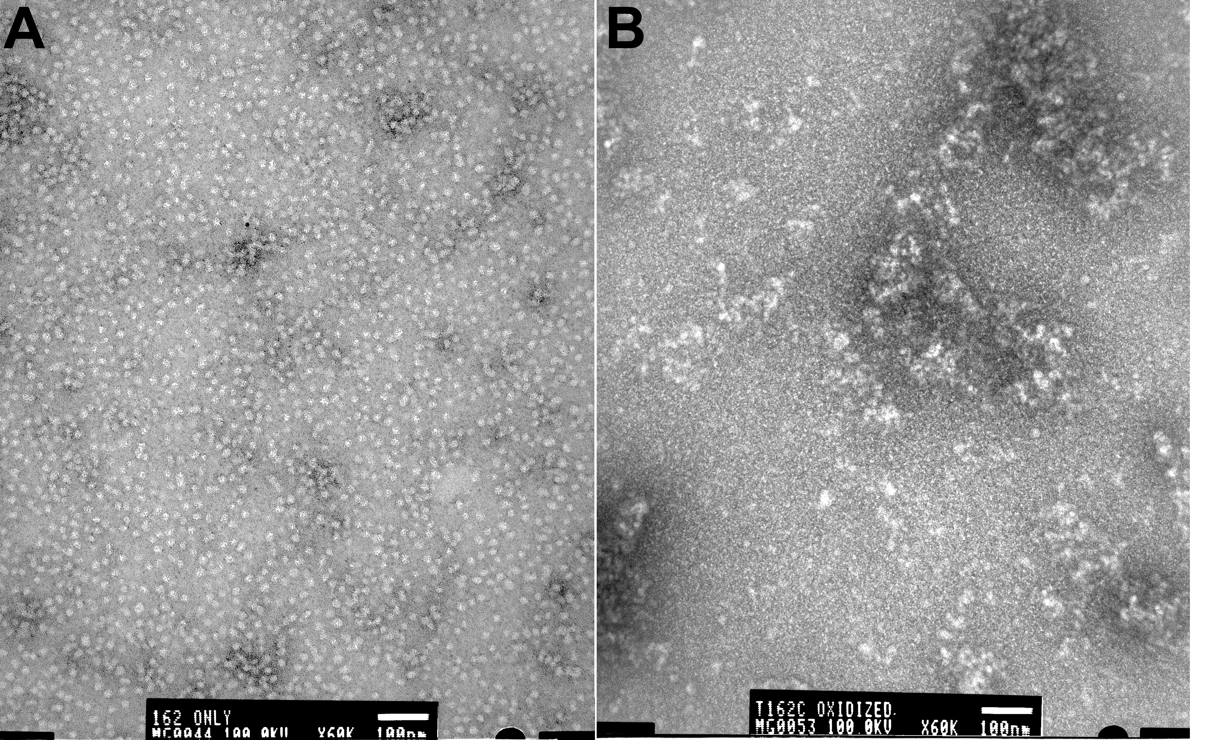
Figure 4. Electron microscopic image of
αBT162C in reduced and oxidized form. A drop of 1 mg/ml protein was
negatively stained with 2% uranyl acetate and observed under the JOEL
1200EX electron microscope. A: Reduced form shows homogeneous
population of oligomers of 10-15 nm. B: Oxidized αBT162C
forms highly heterogeneous and larger aggregates. The EM analysis
of reduced and oxidized αBT162C confirms the aggregation of
αB-crystallin following oxidation and crosslinking.