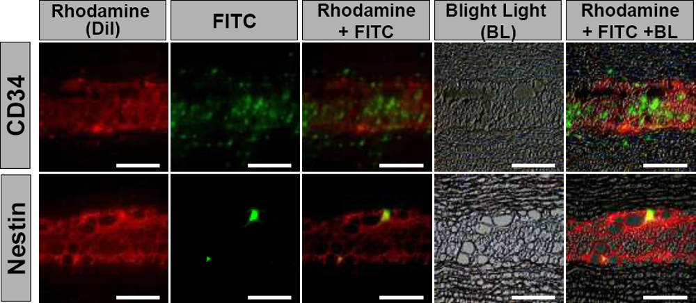
Figure 5. Immunolocalization of
CD34-positive or nestin-positive cells within the transplanted
DiI-positive precursors in the precursor/gelatin group four weeks after
transplantation of gelatin hydrogels with corneal fibroblast
precursors. Bright light (black and white, background), rhodamine (red,
the transplanted DiI-labeled corneal fibroblast precursors in the
gelatin hydrogels), and FITC (green, CD34- or nestin-positive cells)
are superimposed with Adobe Photoshop software. Whole transplanted
gelatin hydrogels are shown in light red by many DiI-positive corneal
fibroblast precursors. A few CD34-positive cells or nestin-positive
spindle cells are scattered within the gelatin hydrogels. Scale bar=100
μm.