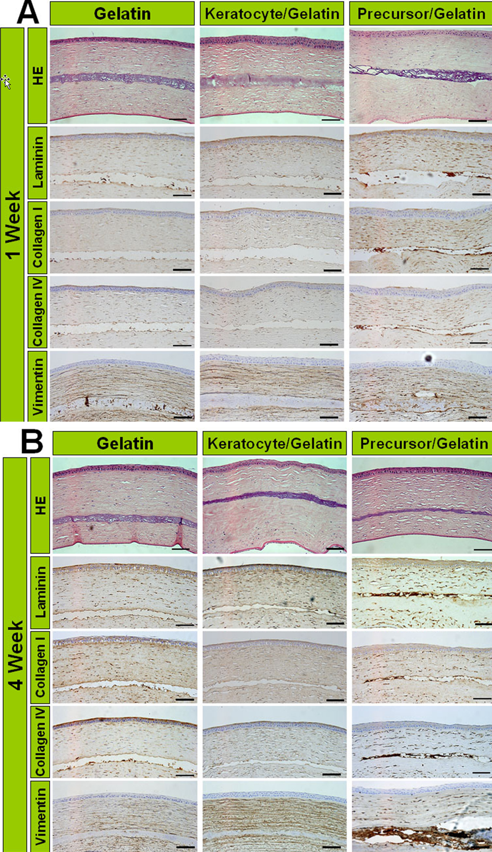
Figure 4. Histological findings and
immunocytochemical analysis of extracellular matrix at one week and
four weeks after transplantation. The transplanted gelatin hydrogels
are found in the corneal stroma in all groups. H&E staining reveals
no mononuclear cell infiltration around gelatin hydrogels in all
groups. The precursor/gelatin group shows more intense staining of
laminin, type I collagen, type IV collagen, and vimentin in the
transplanted gelatin than in the gelatin and fibroblast/gelatin groups
one week after transplantation (A). These expressions in the
precursor/gelatin group increase four weeks after transplantation (B).
Scale bar=100 μm.