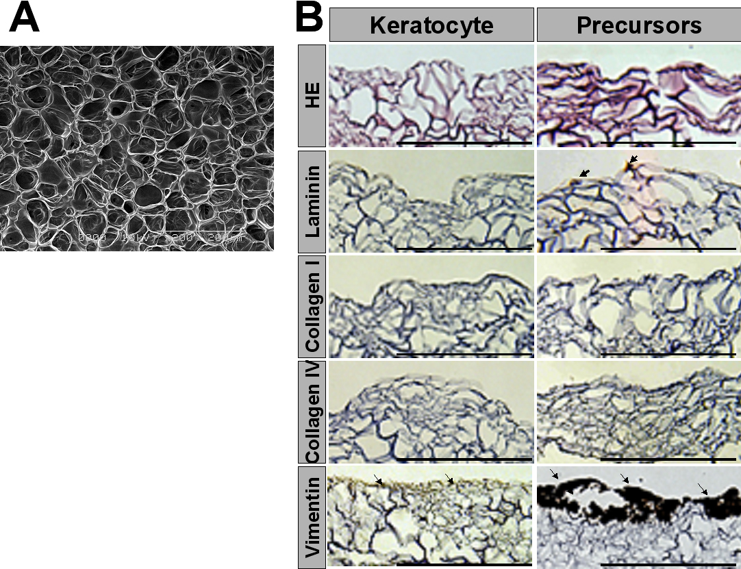
Figure 3. Immunohistochemical analysis of
extracellular matrix in porous gelatin hydrogels with corneal
fibroblasts or fibroblast precursors. Corneal fibroblasts or fibroblast
precursors were seeded onto porous gelatin hydrogels and cultured for
one week. A: Scanning electron microscopy revealed a porous
structure for the gelatin hydrogels. B: Hematoxylin and eosin
staining and immunohistochemical analysis of vimentin and ECM in porous
gelatin hydrogels seven days after seeding of corneal fibroblasts or
fibroblast precursors are shown. Vimentin staining was more intense in
the gelatin hydrogels with corneal fibroblast precursors than in those
with corneal fibroblasts (arrows). Other ECM components such as
laminin, type I collagen, and type IV collagen are not expressed in the
gelatin hydrogels with corneal fibroblasts or fibroblast precursors
before transplantation except for a weak expression of laminin in the
gelatin hydrogels with corneal fibroblast precursors (arrow). Scale
bar=200 μm in B.