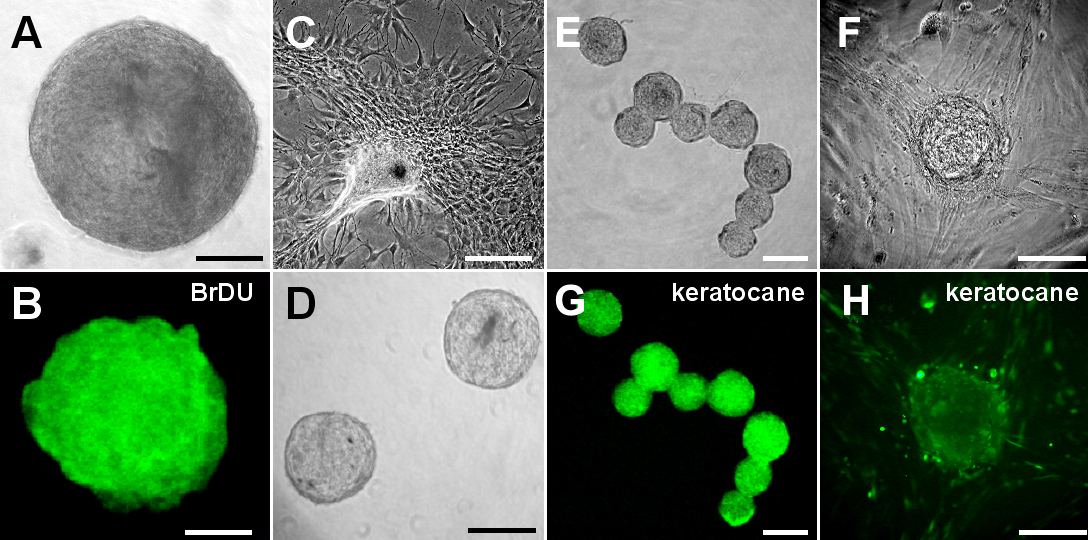
Figure 2. Sphere formation from rabbit
corneal fibroblasts. A: A representative day 7 sphere from
rabbit corneal fibroblasts had a diameter of approximately 300 μm. B:
Each primary sphere was positive for BrdU on day 7. C: The
differentiated progeny from the primary sphere showed a typical
fibroblast-like morphology. D: Secondary spheres were generated
from dissociated primary spheres. E and F: Day 7
spheres were positive for keratocan. G and H: Progenies
derived from the sphere are positive for keratocan. Scale bar=100 μm.