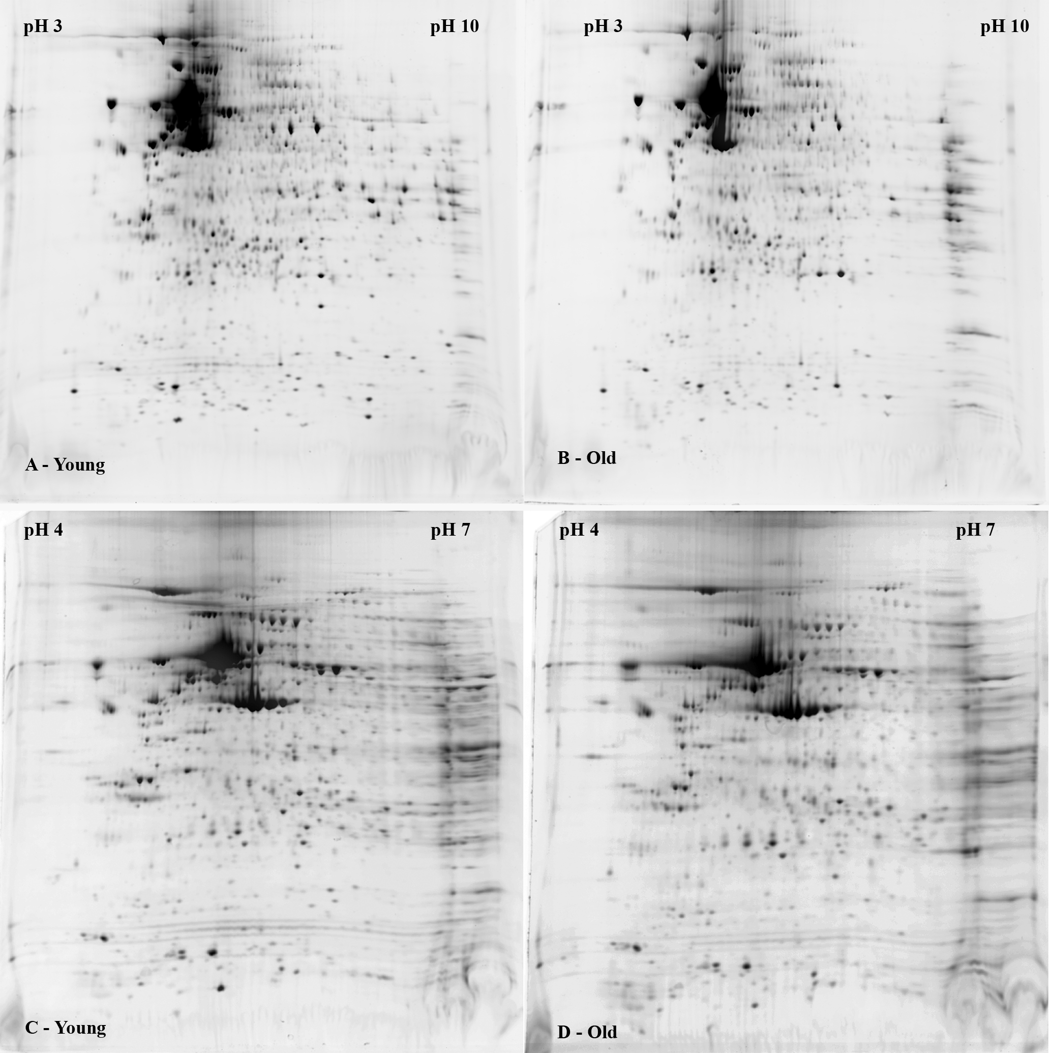
Figure 2. Representative two-dimensional
gel images showing separation of proteins extracted from human corneal
endothelial cells cultured from young and older donors. Extracted
protein was pooled from five young (<30 years old; A,C)
and five older donors (>50 years old; B,D). Equal
amounts of protein were separated on either pH 3–10 IPG strips (A,B)
or pH 4–7 IPG strips (C,D) followed by separation on
8%–16% polyacrylamide gels. Protein spots were stained with SyproRuby.
Images were obtained using a ProEXPRESS Proteomic Imaging System.