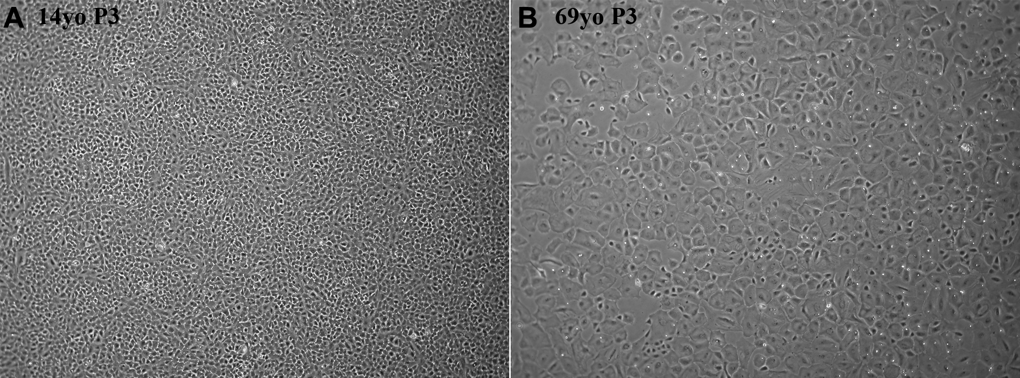
Figure 1. Representative phase-contrast
images of confluent passage 3 human corneal endothelial cells cultured
from a 14-year-old donor and a 69-year-old donor. HCEC from these
donors were among those used for subsequent proteomic analysis. Cells
cultured from older donors were consistently larger and displayed more
variable shapes than cells from young donors. Original magnification:
4X.