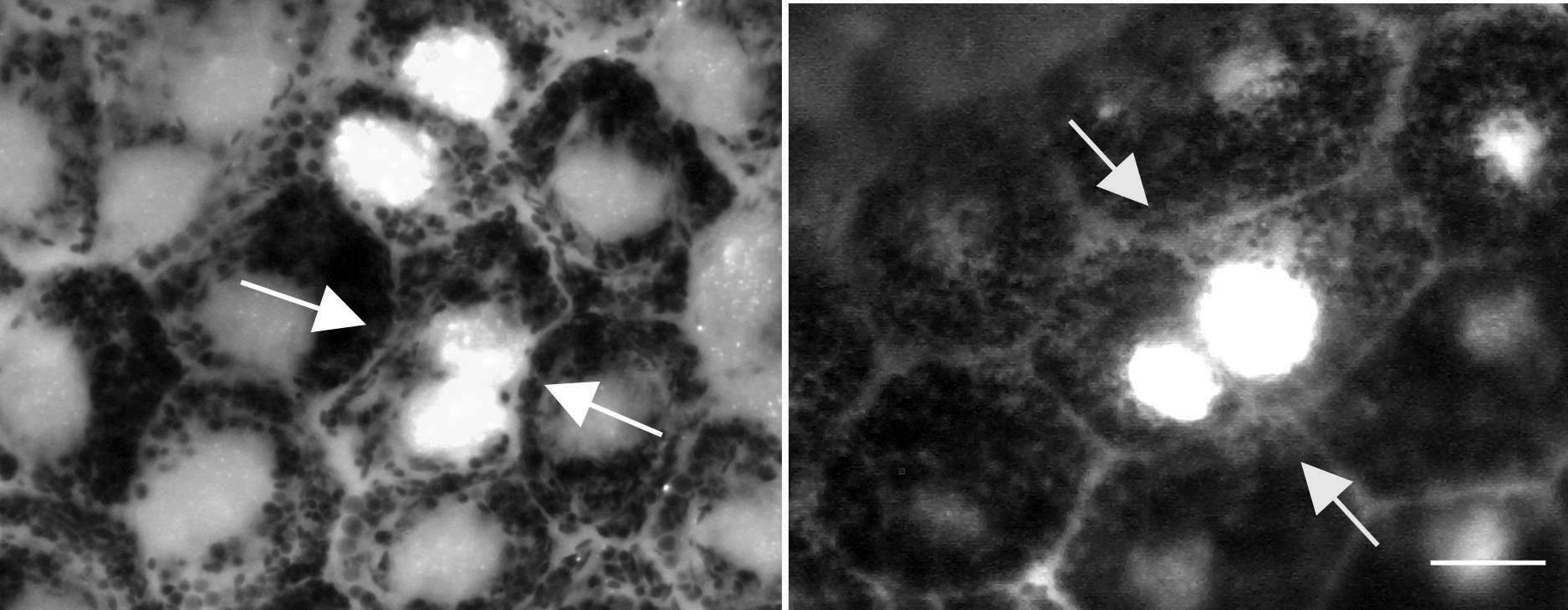
Figure 5. RPE cells were identified that
appear to be going through full cell division. In the peripheral
retinal regions, a small number of cells could be identified that
appeared to be passing through full cell division. In both pictures,
arrows point to two labeled nuclei that appear to be forming a plasma
membrane between them. Both sets of cells appear irregular in the RPE
cell matrix. Taken together with the finding that there was no increase
in the number of binucleated cells in the peripheral retina, these
photographs demonstrate that at least some of the cells in this region
were undergoing full cell division.