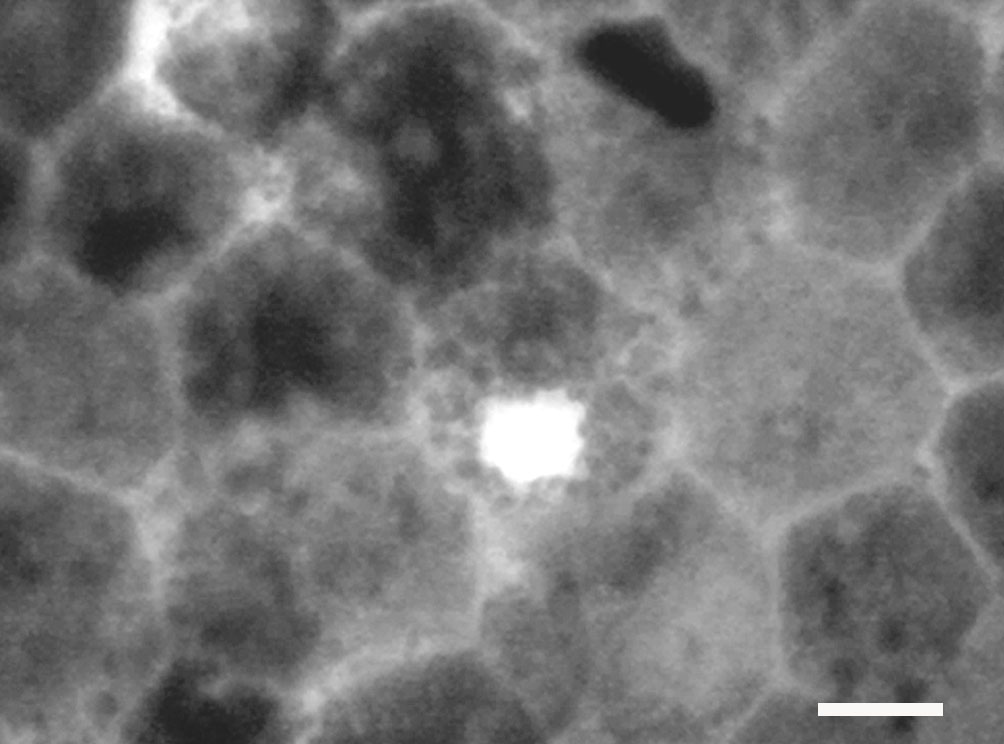
Figure 3. Ki67-positive cells in the
sample of human eye tissue. Only a small strip of tissue, spanning from
the equatorial to peripheral regions, was examined. While Ki67-positive
cells were clearly present, it was not possible to estimate the number
of these cells or map their retinal location. Scale bar equals 10 µm.