Figure 1. Immunohistochemical staining of p16INK4a,
p21WAF1/CIP1, p27KIP1, and p53. Representative
fresh-frozen sections of human corneas from donors at various ages (A:
p16, 18 years; B: p21, 33 years; C: p27, 54 years; D:
p53, 18 years; E: control, 68 years) are shown. Positive
staining for all four senescence-related genes are clearly visible in
the endothelial nuclei. Magnification; 400×.
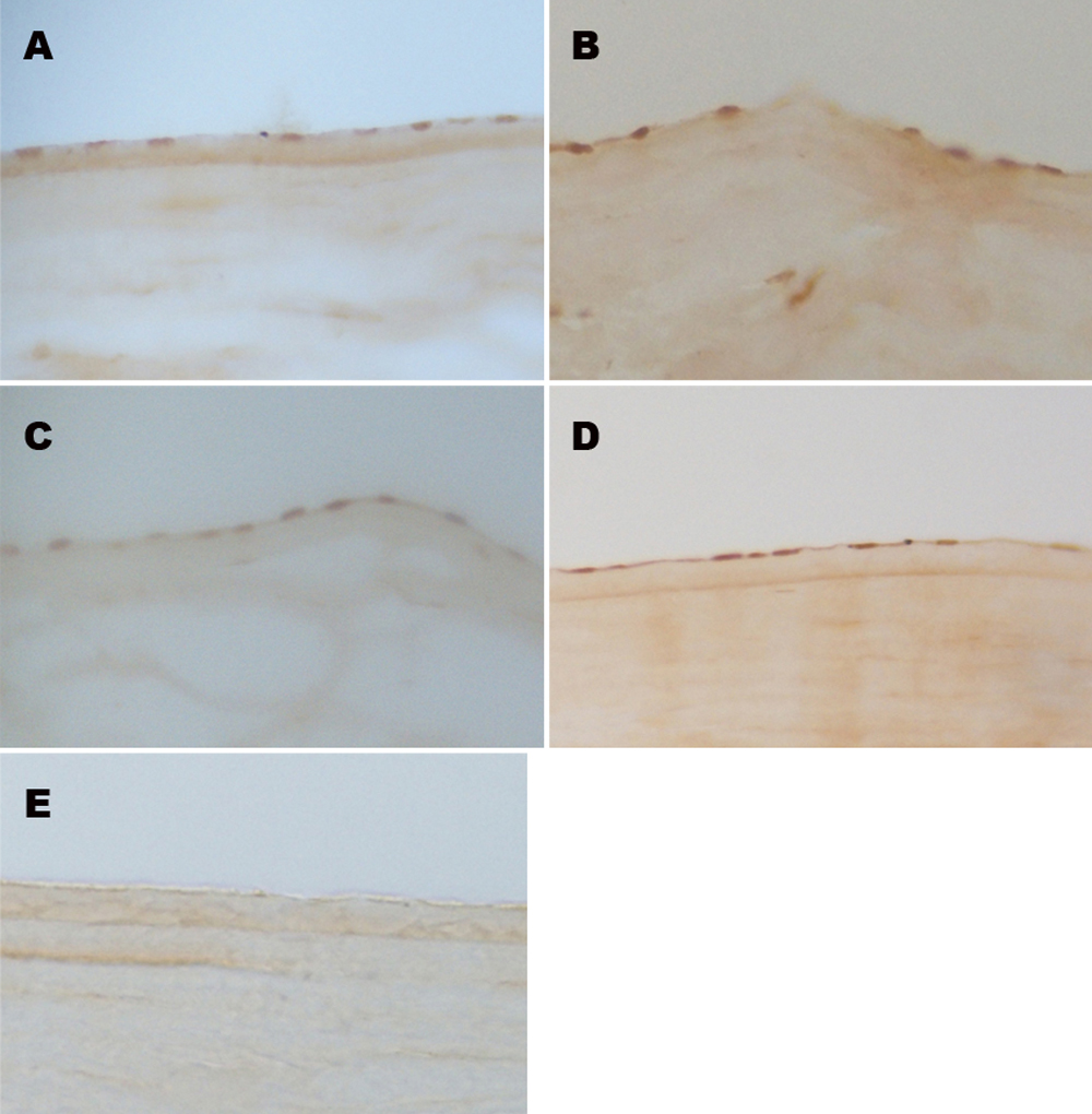
 Figure 1 of Song, Mol Vis 2008; 14:161-170.