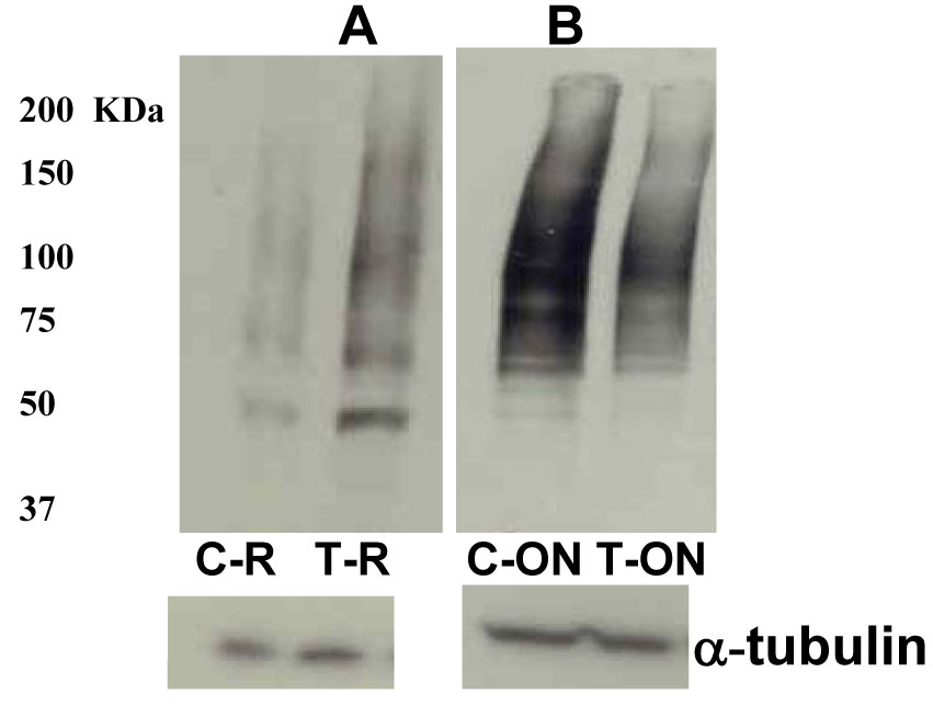
Figures 9. Elevation of IOP increased
ubiquitination in retinal extracts and decreased ubiquitination in
optic nerve extracts. Ubiquitination starts with monoubiquitination at
multiple lysine residues then extending the ubiquitin chains by the
sequential addition of multiple ubiquitin moieties and that is why when
using the anti-ubiquitin antibodies, immunoreactive smear is detected
that reflects the covalent attachment of multiple ubiquitin chains to
individual protein molecule. Western blot with anti-ubiquitin
antibodies revealed a characteristic ubiquitin-positive smear in each
lane corresponding to the amount of ubiquitination on proteins, and
elevation of IOP enhanced ubiquitination in retinal extracts (A)
while decreased ubiquitination in optic nerve extracts (B).
Abbreviations: C-R is control retina, and T-R is elevated IOP retina;
C-ON is control optic nerve, and T-ON is elevated IOP optic nerve.