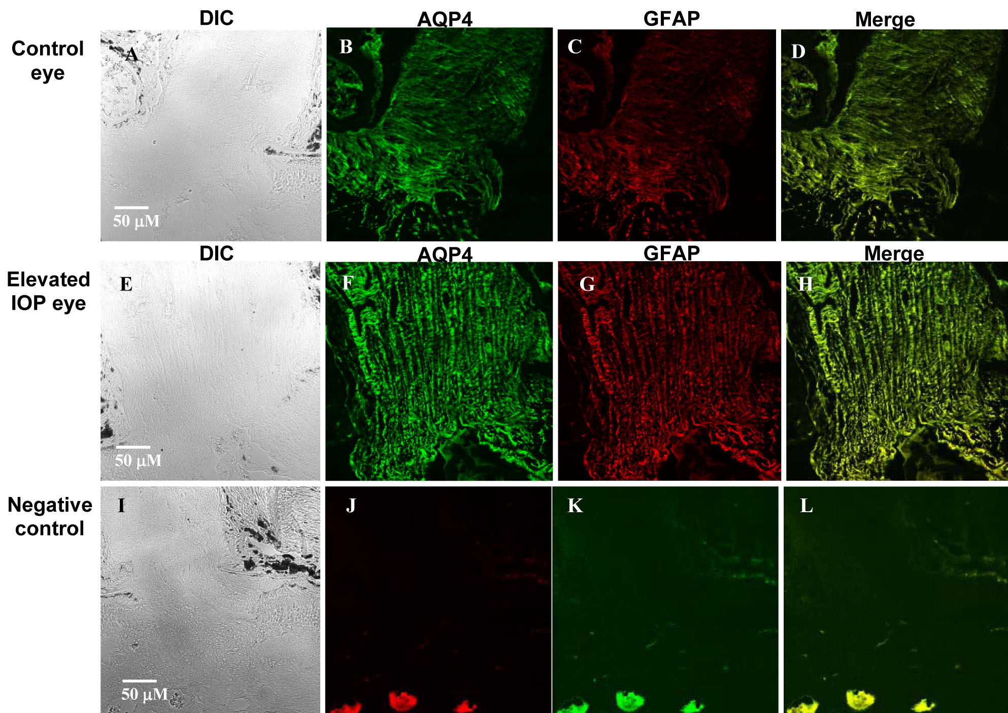
Figure 7. Expression of immunoreactive
AQP4 (green) and glial fibrillary acidic protein (GFAP; red) in the
optic nerve of rats exposed to experimentally elevated intraocular
pressure. Elevation of IOP was performed as described by Morrison et al
[
22]. The
contralateral (control) eyes showed modest AQP4 and GFAP expression (
B-D).
In contrast, AQP4 and GFAP labeling at the optic nerve was consistently
more intense in eyes exposed to elevated IOP compared to control eyes (
F-H).
A representative figure of the optic nerve is shown for one rat exposed
to >500 mmHg days of IOP elevation.
A and
E are DIC
images,
B and
F represent AQP4 labeling,
C and
G
represent GFAP labeling, and
D and
H represent merged
images of AQP4 and GFAP labeling (yellow). Scale bars represents 50 μm.
Staining was performed without primary antibodies (
I-L) that
shows no labeling and was used as a negative control.