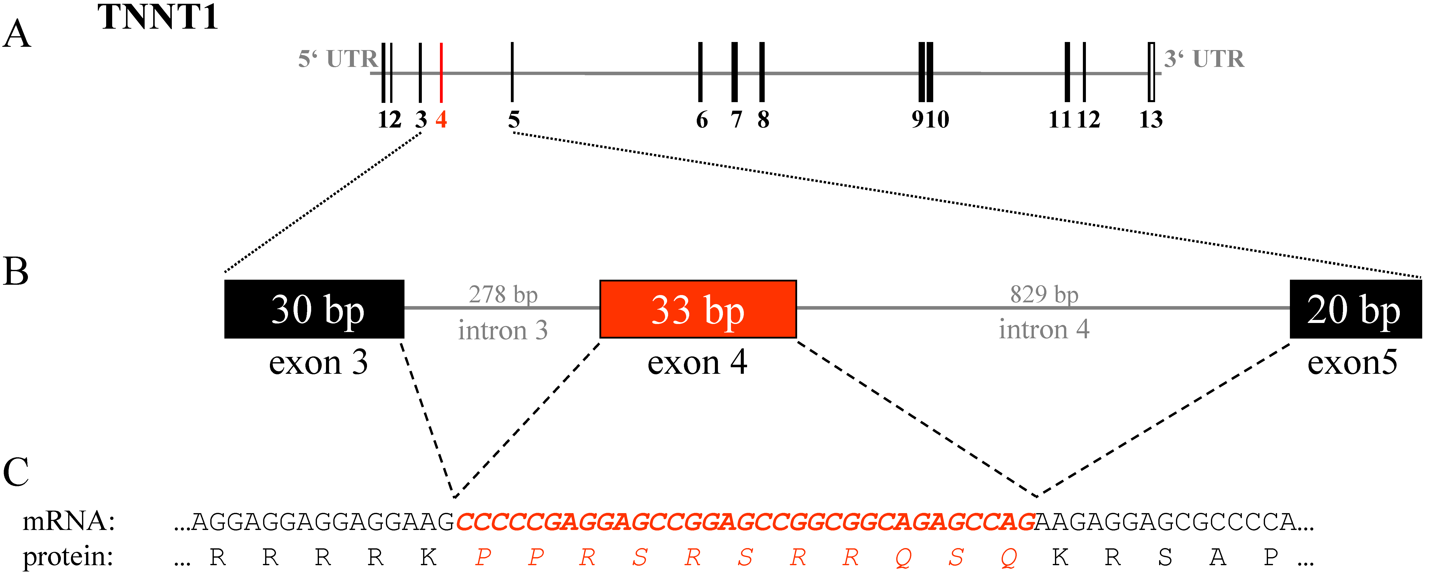
Figure 4. Splice variant of TNNT1.A:
The genomic structure of TNNT1 is shown. B: Detailed
illustration of the genomic region from exon 3 to exon 5 is given. C:
The mRNA sequence of exon 4, their flanking sequences, and the
corresponding translated amino acids are pictured. Translated exons are
shown as solid boxes. Untranslated regions are shown as gray bars. The
exons are numbered by Arabic numerals. The length in base pairs is
given for each sequence. Exon 4 is marked in red. The corresponding
mRNA sequence and the translation product of exon 4 are printed in italics.