Figure 2 of
Szaflik, Mol Vis 2008; 14:1713-1718.
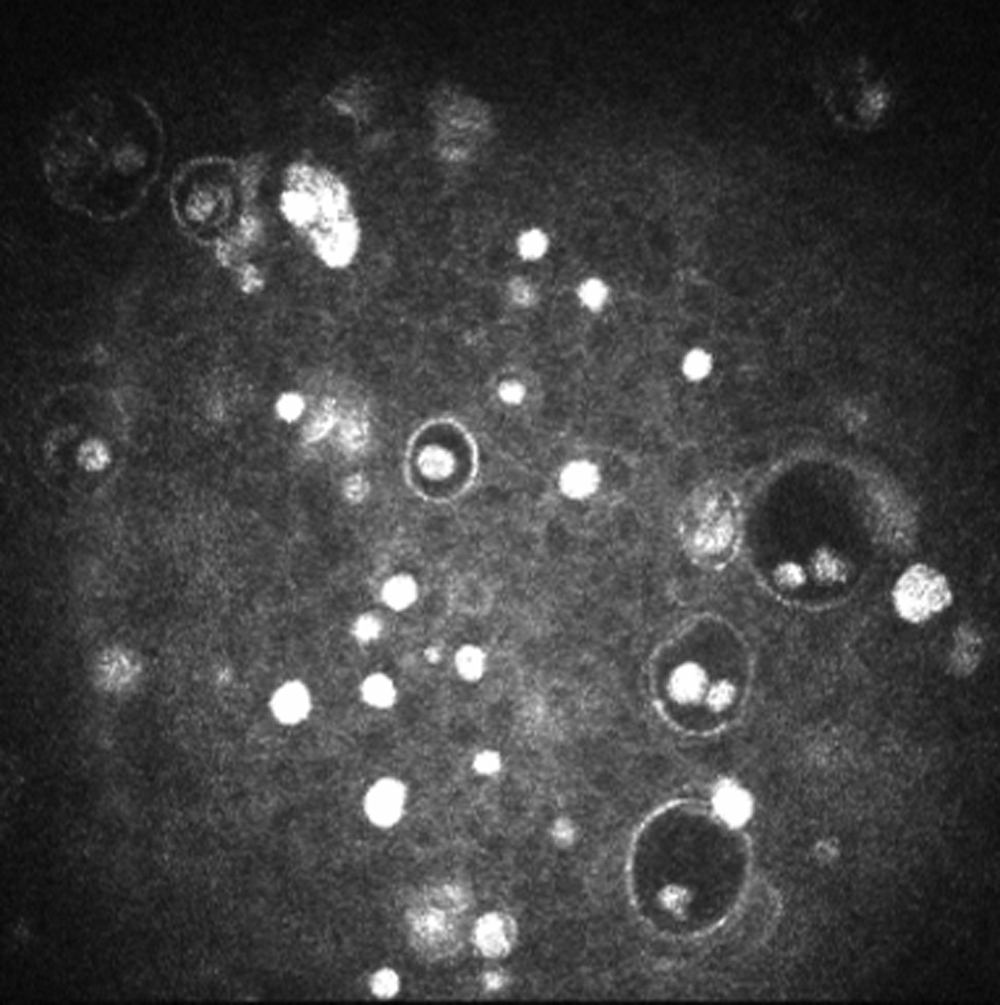
Figure 2.
Confocal microscopy image of proband’s cornea. This image shows the presence of hyperreflective material within the intraepithelial cysts.