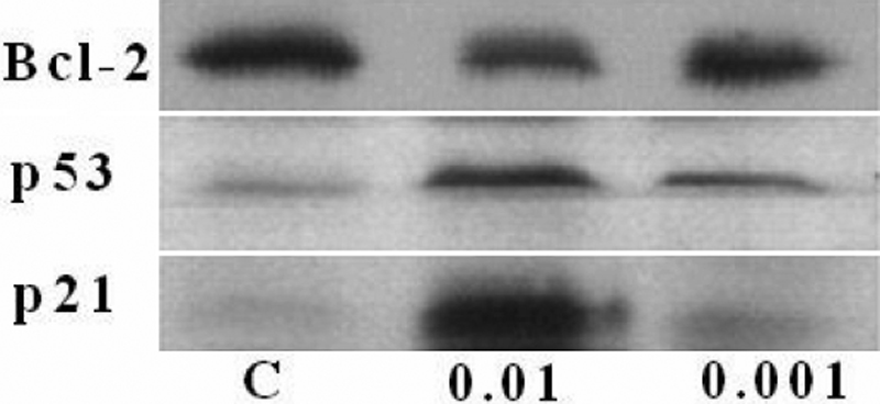
Figure 6. Western blot assay of proteins
involved in apoptosis in corneal endothelial cells. Following
incubation of cells either in the absence of mitomycin C for 24 h
(labeled as C in the image) or in the presence of 0.01 mg/ml (labeled
as 0.01 in the image) and 0.001 (labeled as 0.001 in the image) mg/ml
mitomycin C for 24 h, cells were subjected to SDS–PAGE electrophoresis
and immunoblotting using antisera against Bcl-2, p53, and p21.
Densitometric analysis of protein bands showed the optical density
values of various proteins with a detailed description in the Results.
The respective control values of three proteins were assumed as 100%
response. The optical density of the Bcl-2 protein decreased to 64%±4%
at 0.01 mg/ml and 86%±5% at 0.001 mg/ml in comparison with the control
protein value of 100%±4%. The p53 protein only significantly increased
at 0.01 mg/ml to 139%±4% over the control value. The p21 protein
increased to 373%±6% at 0.01 mg/ml and 149%±7% at 0.001 mg/ml in
comparison with the control value. Two other independent experiments
produced similar results.