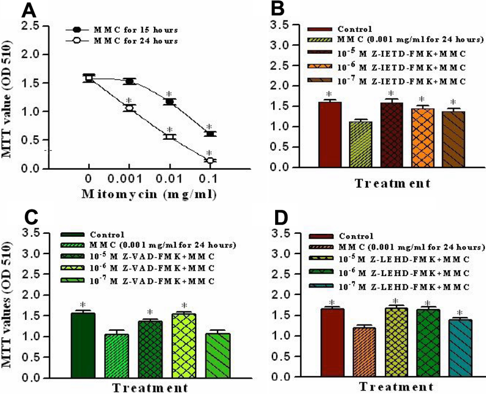
Figure 1. Effects of mitomycin C on cell
viability and apoptotic caspase pathways. A: Cell viability was
measured under exposure to various concentrations of mitomycin C at 15
h and 24 h in cultured porcine corneal endothelial cells. B, C,
and D: The mitomycin-induced apoptotic caspase pathways were
identified with different caspase inhibitors. The cells were pretreated
with a caspase-8 inhibitor (Z-IETD-FMK; B), a general caspase
inhibitor (Z-VAD-FMK; C), or a caspase-9 inhibitor (Z-LEHD-FMK;
D) for 1 h and then exposed to 0.001 mg/ml mitomycin C for 24 h.
Data are presented as means±SD from six replicates and three
independent experiments. The asterisk denotes that p<0.05 when
comparing the control group in A with the MMC only treated
group in B, C, and D.