Figure 7. Representative confocal micrographs of various tissues 6 h after periocular administration of 20 nm particles. Following periocular
administration of 400 µg dose of 20 nm particles to live rats, the nanoparticles can be found in the organs of the reticulo-endothelial
system (liver and spleen). The various tissues including the eye, the periocular tissue, the liver and the spleen were removed
and sectioned 6 h after administration. The figure shows the fluorescence images of sections of the: eye (Panels A and B); periocular tissue (Panels C and D); liver (Panels E and F); and spleen (Panels G and H). The left panels (A, C, E, and G) are fluorescence images from control rats that were not dosed with the nanoparticles whereas the right panels (B, D, F, and H) are images from the rats that were dosed with the nanoparticles. Nanoaprticles can be seen in the periocular tissue, spleen
and to some extent in the liver of the dosed animals.
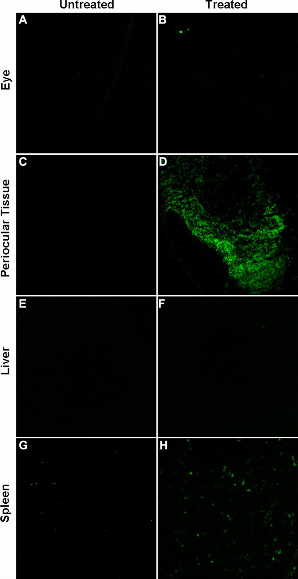
 Figure 7 of
Amrite, Mol Vis 2008; 14:150-160.
Figure 7 of
Amrite, Mol Vis 2008; 14:150-160.