Figure 1. Nanoparticle size distribution
and permeability across sclera and sclera-choroid-RPE. A:
Particle size distribution after 24 h storage in assay buffer used for
transport studies. Majority of the particles are distributed around the
50 nm particle size with another small group of particles of higher
size distribution. B: Particle size distribution after 24 h
storage in assay buffer containing 0.1% tween-20. In the presence of
the surfactant, the particle size distribution shifts towards greater
particle size indicating probable particle aggregation. C:
Transport of nanoparticles (20 nm; 100 µg/ml) across isolated bovine
sclera and sclera-choroid-RPE. The 20 nm particles can cross the sclera
but not the sclera-choroid-RPE combination to any quantifiable extent. D:
Transport of nanoparticles (20 nm; 100 µg/ml) across isolated bovine
sclera and sclera-choroid-RPE in the presence of 0.1% tween-20. The
particle transport across sclera is reduced in the presence of
surfactant probably due to the shift in particle size distribution.
Data are expressed as mean ± s.d. for n = 5-6. No quantifiable
transport was observed either across the sclera or the
sclera-choroid-RPE with the 200 nm particles.
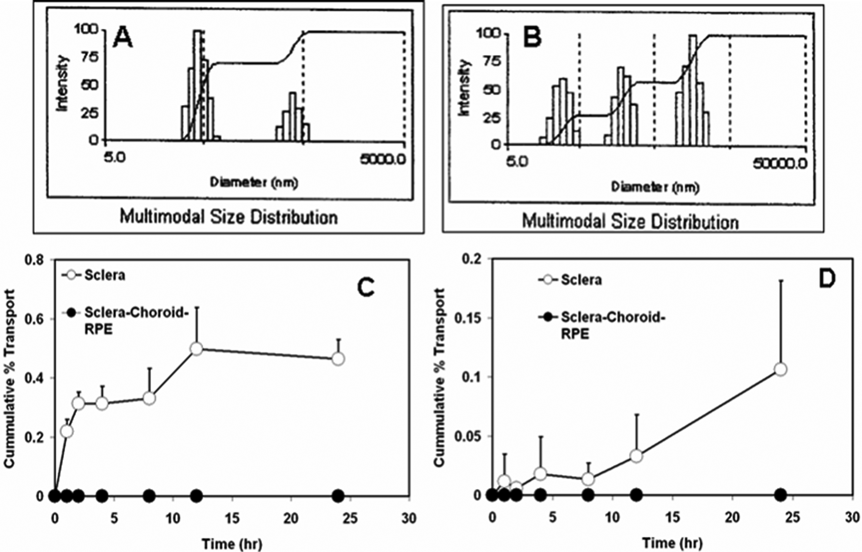
 Figure 1 of Amrite, Mol Vis 2008; 14:150-160.