![]() Figure 5 of
Millar, Mol Vis 2008;
14:10-19.
Figure 5 of
Millar, Mol Vis 2008;
14:10-19.
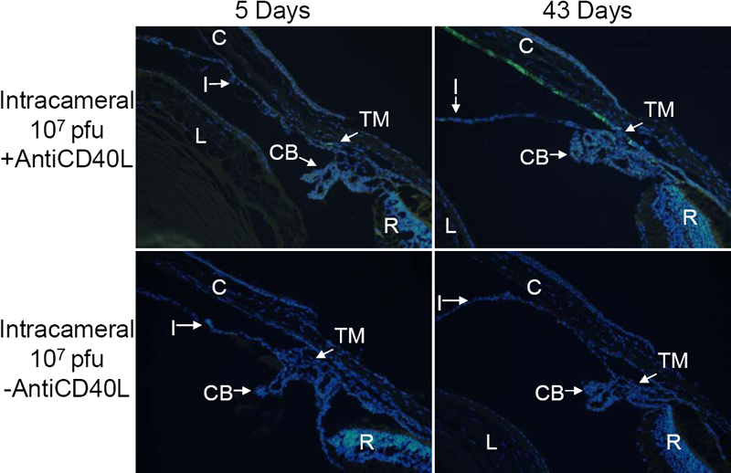
Figure 5. Representative fluorescence photomicrographs of cross-sections of mouse anterior segments treated with intracameral injection of Ad.CMV-GFP (1x10⁷ pfu)
The animals were euthanized at either 5 or 43 days after vector injection with or without anti-CD40L antibody treatment. Green fluorescence = GFP, blue fluorescence = DAPI staining of cell nuclei. The occasional yellowish-green fluorescence represents auto-fluorescence of certain tissues, such as those in the outer nuclear layer and retinal pigmented epithelium, which was also observable in non-injected eyes (Data not shown). Abbreviations: C = cornea; CB = ciliary body; I = iris; L = lens; R = retina; TM = trabecular meshwork.