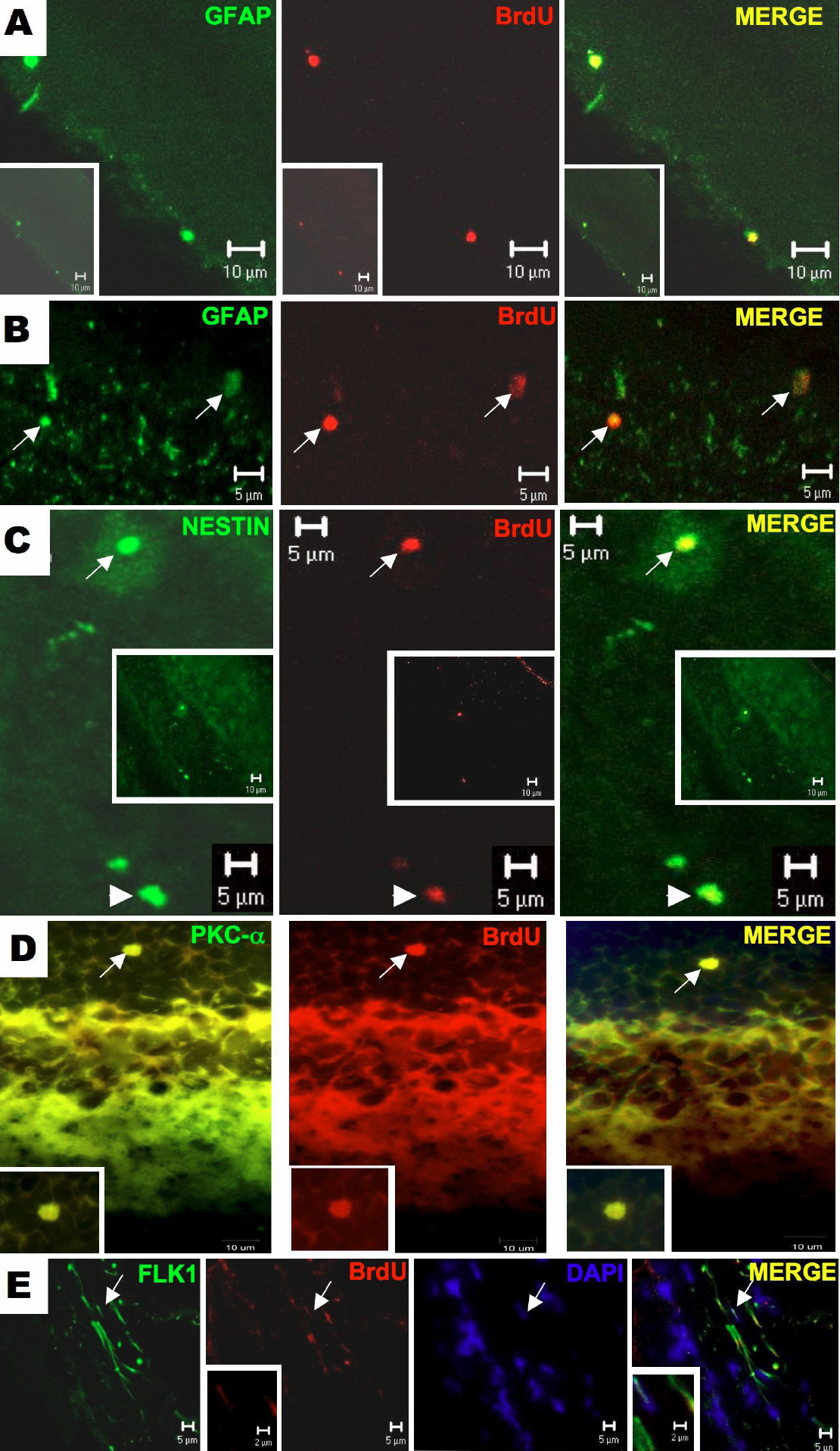
Figure 2. Immunofuorescence assay for
double labeling of BrDU positive cells in retinal sections against
glial, neuron and endothelial markers. In panel A, it is shown
a retinal section showing two cells labeled for glial fibrillar acidic
protein (GFAP) and BrdU in the ganglion cell layer. In B, there
are the presence of two BrdU-positive cells in outer nuclear layer of
the retina co-stained with GFAP antibody. In this slide (C), the
BrDU positive cell is stained for nestin in the inner nuclear (long
arrows) and ganglion cell (short arrows) layers of the retina. For
identification of amacrine/bipolar origin of BrDu positive cell in the
retina, an immunofluorescence for PKC- α antigen was performed. There
is a BrDU positive cell that also stained for PKC- α in the outer
nuclear (D). In panel E, elongated endothelial cells were
identified expressing both Flk-1 and BrdU, localized in theinner
nuclear layer of the retina