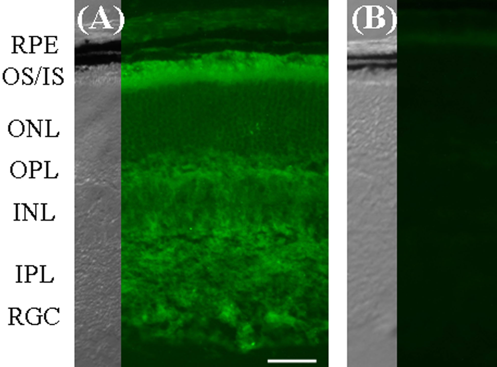
Figure 2. Pla2g7 localization.
Immunohistochemistry was performed in juvenile C57BL/6 (P17) frozen
sections (A), using no primary antibody conditions as the
negative control (B). Pla2g7 was found to be localized
throughout the retina. Relatively elevated levels were found in the
photoreceptor inner and outer segments, whereas moderate staining was
found in the two plexiform layers, as well as the inner nuclear layer
(INL) and the retinal ganglion cell (RGC) layer. For each image, the
corresponding DIC image is provided. The following abbreviations were
used: retinal pigment epithelium (RPE), outer segments (OS), inner
segments (IS), outer nuclear layer (ONL), outer plexiform layer (OPL),
inner nuclear layer (INL), inner plexiform layer (IPL), and RGC:
retinal ganglion cells (RGC). Scale bar in (A) represents 20 μm.