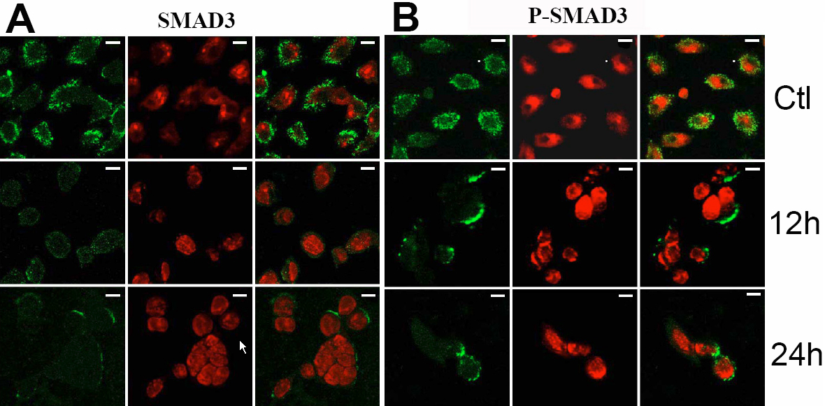
Figure 5. Colocalization of SMAD3 and
phospho-SMAD3 protein in human corneal epithelial cells. FITC marked
the secondary antibody (green; left), and PI dyed the nucleus (red;
middle). Merged images were showed at the right of A and B.
Both SMAD3 (A) and phospho-SMAD3 (B) were more weakly
expressed at 12 h and 24 h p.i. compared to the uninfected cells. Scale
bar: 10 μm.