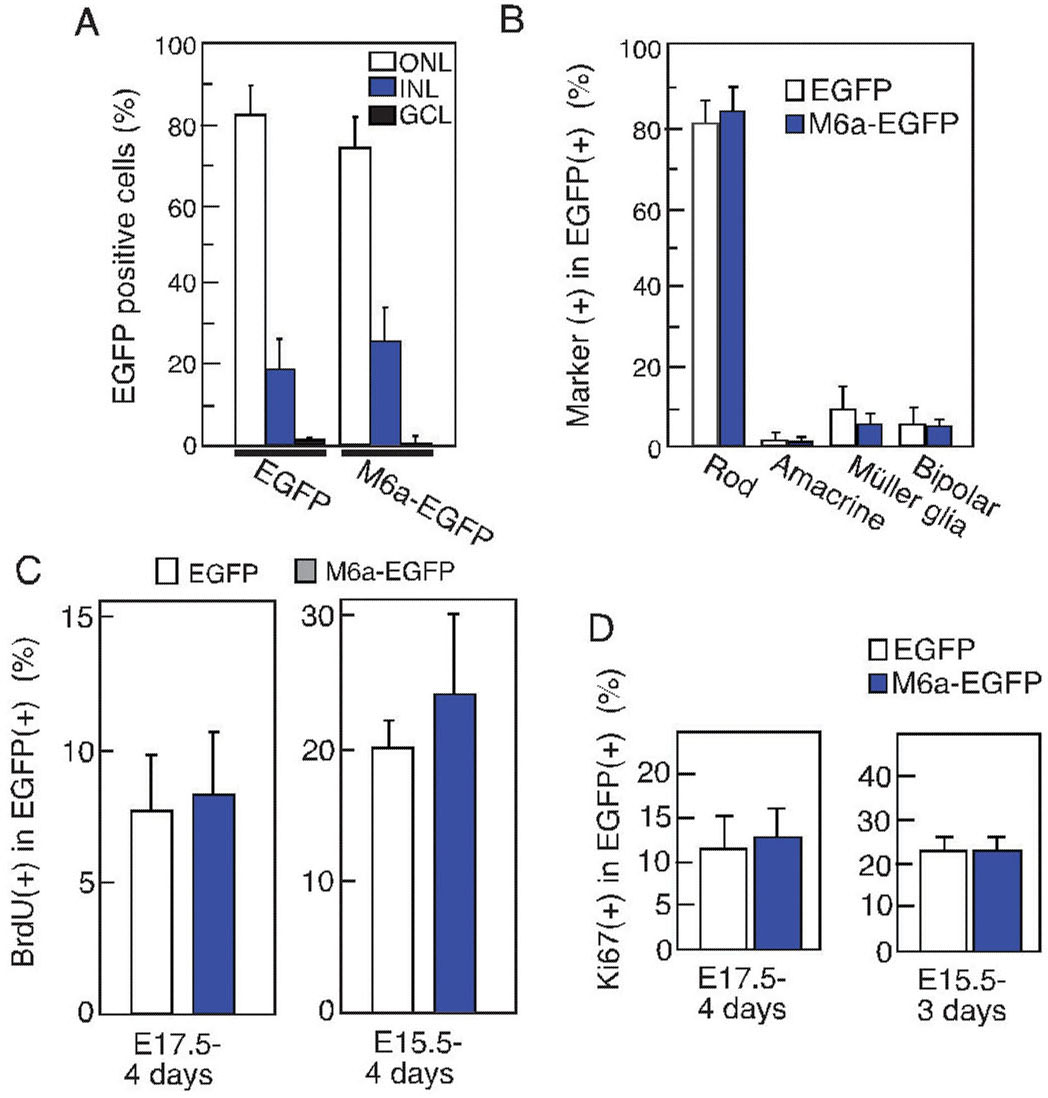
Figure 2. Effects of forced expression of
M6a on retinal differentiation and proliferation. A: Sublayer
distributions of virus-transduced enhanced green fluorescent protein
(EGFP)-positive cells in retinal explants. Retinal explants were
infected with retorovirus particles that encode M6a and EGFP. After 14
days, the explants were harvested and frozen-sections prepared.
Immunostaining was performed using an anti-EGFP antibody. The
percentages of cells in each sublayer are shown. More than 200 cells
were counted for each sample, and the standard deviation (SD) was
calculated from three independent experiments. B:
Differentiation of virus-infected cells examined by immunostaining to
identify subpopulations within the retina. The percentages of
marker-positive cells in the EGFP-positive population are shown.
Rhodopsin for rod, HuC/HuD for amacrine, glutamine synthetase for
Müller glia, and protein kinase C for bipolar were used as markers.
More than 100 cells were examined for each sample, and the average
value from three independent experiments is shown with the SD. C,D:
Proliferation of M6a-expressing retinal cells was examined by measuring
incorporation of bromodeoxyuridine (BrdU; C) or expression of
the Ki67 antigen (D). BrdU was present for the final 24 h of
four days of culture of retinal explants, and frozen sections were
immunostained using antibodies against BrdU. The same samples were
immunostained with the anti-Ki67antibody. The percentage of positive
cells with SD are shown. Listed below each panel is the stage when each
retinal explant was prepared and its culture period. The following
abbreviations are in effect: outer nuclear layer (ONL); inner nuclear
layer (INL); ganglion cell layer (GCL).