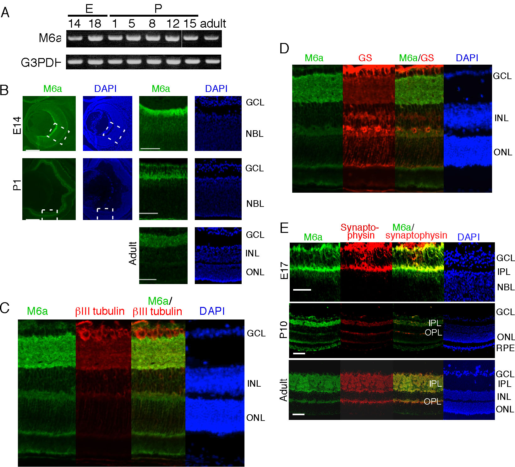
Figure 1. Expression of M6a in mouse
retinas from various developmental stages. A: Semiquantitative
RT–PCR for M6a using total RNA extracted from mouse retinas at various
developmental stages. Glyceraldehyde-3-phosphate dehydrogenase (G3PDH)
was used as the control. B-E: Immunostaining of M6a in frozen
sections of mouse retina from various developmental stages.
Coimmunostaining was performed with anti-M6a and anti-βIII-tubulin (C),
anti-glutamine synthetase (D), or antisynaptophysin (E)
antibodies. The right two columns are magnified figure of the square
area indicated by broken lines in left two columns B. The scale
bar represents 100 μm. The following abbreviations are used in this
figure: inner plexiform layer (IPL); outer plexiform layer (OPL);
ganglion cell layer (GCL); neuroblastic layer (NBL); inner nuclear
layer (INL); outer nuclear layer (ONL); retinal pigment epithelium
(RPE).